 Back
Neurons, Synapses, and Signaling: Study Guide (Campbell Biology Ch. 48)
Study Guide - Smart Notes
 Tailored notes based on your materials, expanded with key definitions, examples, and context.
Neurons, Synapses & Signaling
Overview of Neuron Structure and Function
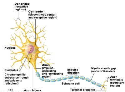
Neurons are specialized cells responsible for transmitting electrical and chemical signals throughout the nervous system. Their structure is closely related to their function in signal conduction and communication.
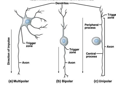
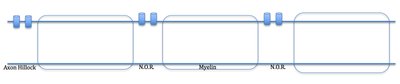
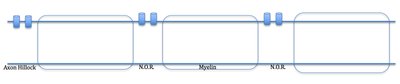
Multipolar neurons have multiple dendrites and a single axon; the axon hillock is the trigger zone for action potential (AP) generation.
Bipolar and unipolar neurons lack a distinct axon hillock but possess trigger zones rich in voltage-gated (VG) ion channels.
Trigger zone: Region where AP is first generated due to high density of VG ion channels.
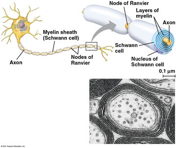
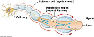
Myelin sheath: Insulating layer formed by Schwann cells, increases conduction velocity.
Nodes of Ranvier (NOR): Gaps in myelin sheath with high concentration of VG ion channels.
Propagation and Conduction of Action Potentials
Action potentials are self-propagating electrical signals that travel along the axon. Their conduction is influenced by myelination and the presence of NOR.
AP is initiated at the trigger zone and travels unidirectionally toward synaptic terminals.
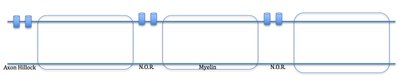
In myelinated neurons, AP propagation involves local flow of depolarizing current (graded potential) that rapidly reaches the next NOR.
Myelin prevents charge leakage, allowing the current to travel internally with minimal loss.
AP conduction is one-way due to refractory periods at upstream NOR and axon hillock.
Refractory Periods
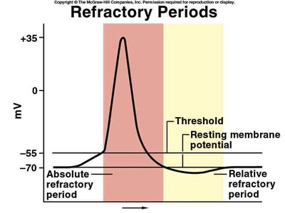
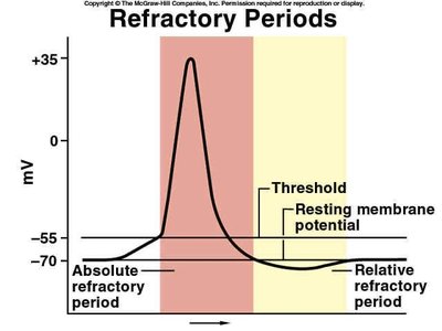
Refractory periods ensure the unidirectional propagation of action potentials and regulate neuronal firing frequency.
Absolute refractory period: No second AP can be initiated; VG channels are locked.
Relative refractory period: Second AP possible only with strong stimulus; VG channels begin to reset.
Saltatory vs. Continuous Conduction
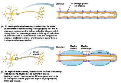
Myelinated neurons conduct APs much faster than unmyelinated neurons due to saltatory conduction.
Saltatory conduction: AP appears to jump from one NOR to the next, increasing speed (up to 30x faster).
Continuous conduction: In unmyelinated neurons, AP is regenerated along the entire axon, resulting in slower transmission.
Wider axon diameter also increases conduction velocity.

Synapses: Neuronal Communication
Types of Synapses
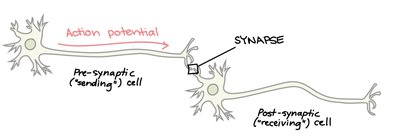
Neurons communicate via synapses, which are either electrical or chemical. Chemical synapses are predominant in the nervous system.
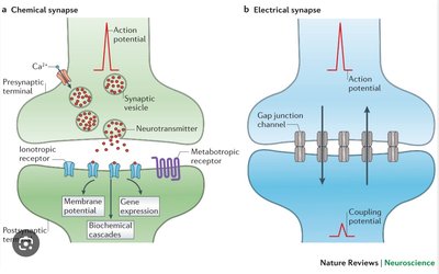
Electrical synapses: Direct ion flow via gap junctions; rapid transmission.
Chemical synapses: Neurotransmitter (NT) release mediates signal transmission.
Events at the Chemical Synapse
Signal transmission at chemical synapses involves a series of steps leading to NT release and postsynaptic response.
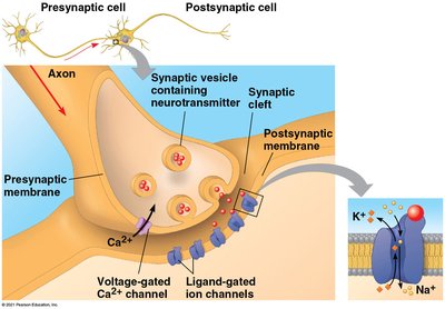
AP reaches axon terminal.
VG-Ca2+ channels open; Ca2+ influx occurs.
Ca2+ influx triggers synaptic vesicles to release NTs via exocytosis.
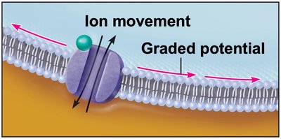
NTs diffuse across synaptic cleft (~50 nm) and bind to ligand-gated ion channels on postsynaptic membrane, causing a graded potential.
Ligand-Gated Ion Channels
Ligand-gated (ionotropic) channels open in response to NT binding, allowing specific ions to flow and alter membrane potential.
Ligand: Molecule that binds to a receptor, triggering a biological response (e.g., NTs, hormones).
Ion flow produces a graded potential in the postsynaptic cell.
Postsynaptic Potentials: EPSP and IPSP
The postsynaptic response depends on the type of ion channel opened by NT binding.
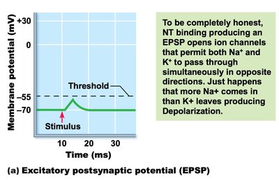
Excitatory Postsynaptic Potential (EPSP): NT opens Na+ channels, causing depolarization and moving membrane toward threshold.
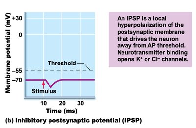
Inhibitory Postsynaptic Potential (IPSP): NT opens K+ or Cl- channels, causing hyperpolarization and moving membrane away from threshold.
Summation of Postsynaptic Potentials
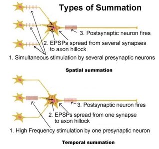
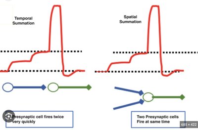
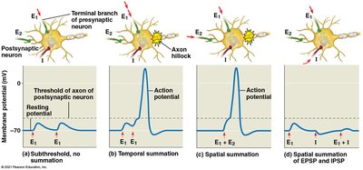
Multiple postsynaptic potentials can combine to produce a larger effect, potentially triggering an AP.
Spatial summation: Multiple presynaptic neurons stimulate a postsynaptic neuron simultaneously.
Temporal summation: One presynaptic neuron fires repeatedly, increasing NT release.
EPSPs and IPSPs can cancel each other out, affecting AP generation.
Termination of Synaptic Signaling
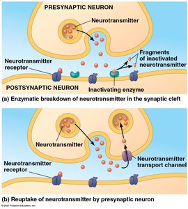
Synaptic signaling is terminated by removal of excess NTs from the synaptic cleft.
NTs are removed via enzymatic hydrolysis or reuptake by the presynaptic neuron.
This ensures precise control of synaptic transmission and prevents continuous stimulation.
Summary Table: EPSP vs. IPSP
Type | Ion Channel Opened | Effect on Membrane Potential | Outcome |
|---|---|---|---|
EPSP | Na+ (sometimes both Na+ and K+) | Depolarization | Moves membrane toward threshold, increases AP likelihood |
IPSP | K+ or Cl- | Hyperpolarization | Moves membrane away from threshold, decreases AP likelihood |
Key Equations
Membrane potential change (graded potential):
Threshold for action potential:
Resting membrane potential:
Additional info: The notes above expand on the original slides with definitions, context, and examples to ensure completeness and clarity for exam preparation.
