Back
BackNeurons, Synapses, and Signaling: Study Notes
Study Guide - Smart Notes
 Tailored notes based on your materials, expanded with key definitions, examples, and context.
Neurons, Synapses, and Signaling
Overview of Neuronal Communication
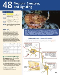
Neurons are specialized cells that transmit information throughout the nervous system using electrical and chemical signals. This chapter explores the structure and function of neurons, the mechanisms underlying signal transmission, and the role of synapses in communication between cells.
Neurons are the basic units of the nervous system, responsible for receiving, processing, and transmitting information.
Synapses are junctions where neurons communicate with other neurons or effector cells.
Signaling involves both electrical impulses (action potentials) and chemical messengers (neurotransmitters).
Neuron Structure and Function
Organization and Information Transfer
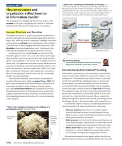
The structure of a neuron is closely related to its function in information transfer. Neurons consist of a cell body, dendrites, and an axon. Dendrites receive signals, while the axon transmits signals to other cells.
Cell body (soma): Contains the nucleus and organelles.
Dendrites: Branch-like extensions that receive signals from other neurons.
Axon: Long projection that transmits electrical impulses away from the cell body.
Axon terminals: Release neurotransmitters to communicate with other cells.
Types of Neurons
Neurons are classified based on their function:
Sensory neurons: Transmit information from sensory receptors to the central nervous system.
Interneurons: Connect neurons within the central nervous system and integrate information.
Motor neurons: Transmit signals from the central nervous system to muscles or glands.
Ion Pumps, Ion Channels, and the Resting Potential
Establishing the Resting Potential
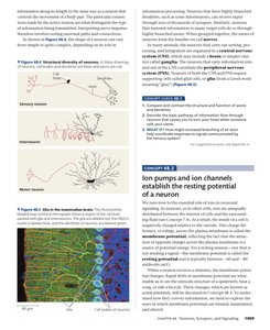
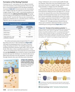
The resting potential of a neuron is the electrical potential difference across the plasma membrane when the cell is not transmitting a signal. It is primarily established by ion pumps and channels.
Sodium-potassium pump (Na+/K+ ATPase): Actively transports 3 Na+ ions out and 2 K+ ions into the cell, maintaining concentration gradients.
Ion channels: Allow passive movement of ions across the membrane, contributing to the membrane potential.
Resting potential: Typically around -70 mV in neurons.
Ion | Inside (mM) | Outside (mM) |
|---|---|---|
K+ | 140 | 5 |
Na+ | 15 | 150 |
Cl- | 10 | 120 |
Protein- | 100 | 2 |
Modeling the Resting Potential
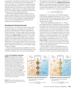
The Nernst equation is used to calculate the equilibrium potential for a particular ion:
Nernst equation:
Where R is the gas constant, T is temperature, z is the charge of the ion, and F is Faraday's constant.
Action Potentials
Signals Conducted by Axons
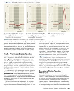
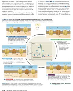
Action potentials are rapid changes in membrane potential that travel along the axon, allowing neurons to transmit signals over long distances.
Depolarization: Membrane potential becomes less negative due to Na+ influx.
Repolarization: Membrane potential returns to resting state due to K+ efflux.
Threshold: Minimum depolarization required to trigger an action potential.
All-or-none response: Action potentials occur fully or not at all.
Generation and Propagation of Action Potentials
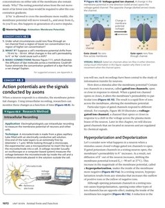
Voltage-gated ion channels play a critical role in the generation and propagation of action potentials.
Voltage-gated Na+ channels: Open rapidly during depolarization, allowing Na+ influx.
Voltage-gated K+ channels: Open more slowly, allowing K+ efflux during repolarization.
Refractory period: Time during which a neuron cannot fire another action potential.
Conduction of Action Potentials
Mechanisms of Conduction
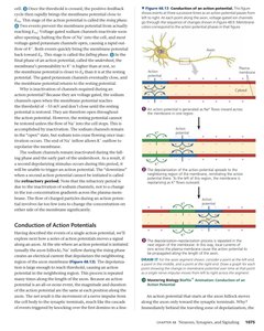
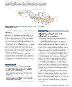
Action potentials are conducted along the axon by the sequential opening of voltage-gated ion channels. In myelinated axons, conduction is faster due to saltatory conduction.
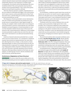
Saltatory conduction: Action potentials jump from one node of Ranvier to the next in myelinated axons.
Myelin sheath: Insulating layer that increases the speed of signal transmission.
Nodes of Ranvier: Gaps in the myelin sheath where ion channels are concentrated.
Synaptic Transmission
Neurons Communicate at Synapses
Neurons communicate with other cells at synapses, which can be electrical or chemical. Chemical synapses use neurotransmitters to transmit signals across the synaptic cleft.
Presynaptic neuron: Releases neurotransmitters into the synaptic cleft.
Postsynaptic neuron: Receives the signal via neurotransmitter receptors.
Synaptic cleft: Small gap between the presynaptic and postsynaptic cells.
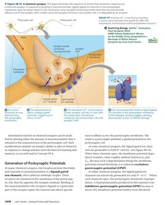
Generation and Summation of Postsynaptic Potentials
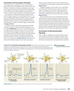
Postsynaptic potentials can be excitatory (EPSP) or inhibitory (IPSP). The summation of these potentials determines whether the postsynaptic neuron will fire an action potential.
Excitatory postsynaptic potential (EPSP): Depolarizes the postsynaptic membrane.
Inhibitory postsynaptic potential (IPSP): Hyperpolarizes the postsynaptic membrane.
Spatial summation: Multiple synapses activate simultaneously.
Temporal summation: Rapid succession of signals at a single synapse.
Termination of Neurotransmitter Signaling
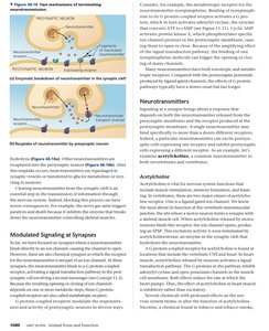
Neurotransmitter signaling is terminated by enzymatic breakdown, reuptake into the presynaptic cell, or diffusion away from the synaptic cleft.
Enzymatic breakdown: Enzymes degrade neurotransmitters in the synaptic cleft.
Reuptake: Neurotransmitters are transported back into the presynaptic neuron.
Neurotransmitters
Types and Functions
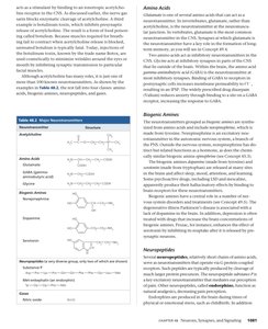
Neurotransmitters are chemical messengers that transmit signals across synapses. They are classified into several major types:
Acetylcholine: Involved in muscle contraction and autonomic nervous system functions.
Amino acids: Includes glutamate (excitatory) and GABA (inhibitory).
Biogenic amines: Includes dopamine, serotonin, and norepinephrine, which regulate mood, arousal, and behavior.
Neuropeptides: Short chains of amino acids that modulate neuronal activity.
Neurotransmitter | Structure |
|---|---|
Acetylcholine | CH3COOCH2CH2N(CH3)3 |
Glutamate | HOOCCH2CH(NH2)COOH |
GABA | NH2CH2CH2CH2COOH |
Dopamine | C8H11NO2 |
Serotonin | C10H12N2O |
Norepinephrine | C8H11NO3 |
Scientific Skills: Data Interpretation
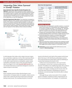
Interpreting Data Values in Scientific Notation
Scientific notation is used to express very large or small values in a concise format, which is essential for analyzing experimental data in neuroscience.
Example: The concentration of neurotransmitters may be expressed as 1.2 × 10⁻⁶ M.
Application: Understanding scientific notation is crucial for interpreting research results and comparing values.
Chapter Review
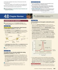
Summary of Key Concepts
Neurons transmit information via electrical and chemical signals.
Ion pumps and channels establish the resting potential.
Action potentials are generated and propagated by voltage-gated ion channels.
Synapses enable communication between neurons and other cells.
Neurotransmitters play diverse roles in neuronal signaling.
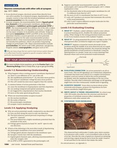
Test Your Understanding
Review the structure and function of neurons.
Explain the mechanisms of action potential generation and propagation.
Describe the role of synapses and neurotransmitters in neuronal communication.