 Back
Neurons, Synapses, Signaling, and the Central Nervous System: Study Guide
Study Guide - Smart Notes
 Tailored notes based on your materials, expanded with key definitions, examples, and context.
Neurons, Synapses, Signaling, and the CNS
Overview of Nervous System Types
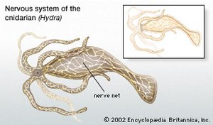
The nervous system is essential for coordinating animal responses to internal and external stimuli. There are two main types: the nerve net and the central nervous system (CNS). The nerve net is a diffuse arrangement of cells, found in cnidarians and ctenophores, while the CNS consists of large clusters of neurons (ganglia) and is characteristic of more complex animals.
Nerve Net: Simple, diffuse network; seen in Hydra and other cnidarians.
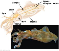
CNS: Aggregated neurons forming brain and spinal cord; enables complex processing.
PNS (Peripheral Nervous System): Neurons outside CNS; connects CNS to muscles, glands, and sensory organs.
Example: Squid with giant axons, used in research on signal transmission.
Neural Pathways and Signal Transmission
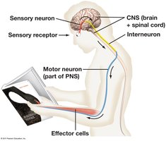
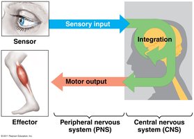
Neural pathways involve sensory receptors, sensory neurons, interneurons, and motor neurons. Sensory input is processed by the CNS, which then sends motor output to effectors (muscles or glands).
Sensory Receptors: Detect environmental changes (e.g., skin, eyes, ears).
Sensory Neurons: Transmit information to CNS.
Interneurons: Connect sensory and motor neurons within CNS.
Motor Neurons: Carry signals from CNS to effectors.
Neuron Structure and Types
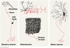
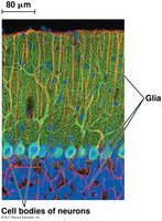
Neurons are specialized cells for signal transmission. They consist of dendrites, a cell body (soma), and an axon. There are three main types: sensory neurons, interneurons, and motor neurons. Glia are supporting cells that nourish and insulate neurons.
Dendrites: Receive signals from other cells.
Cell Body (Soma): Contains nucleus; integrates signals.
Axon: Transmits signals to other neurons or effectors.
Interneurons: Found in CNS; connect sensory and motor neurons.
Glia: Support, nourish, and insulate neurons.
Synapses and Signal Transmission
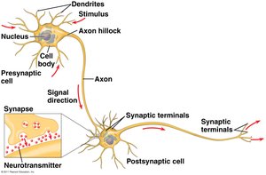
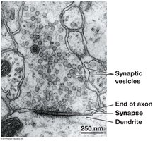
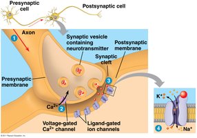
The synapse is the junction between two neurons or between a neuron and an effector cell. The axon terminal releases neurotransmitters, which transmit information across the synaptic cleft to the postsynaptic cell.
Presynaptic Cell: Releases neurotransmitters.
Postsynaptic Cell: Receives signal; can be neuron, muscle, or gland.
Neurotransmitters: Chemical messengers stored in synaptic vesicles.
Electrochemical Basis of Neural Signaling
Membrane Potential and Ion Channels
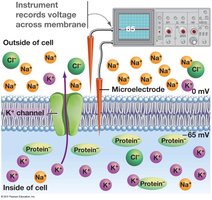
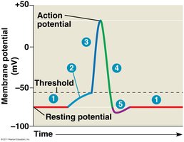
Neurons maintain a membrane potential, an electrical charge difference across the plasma membrane, due to selective permeability and ion gradients. The resting potential is typically -60 to -80 mV.
Membrane Potential: Inside of neuron is negative relative to outside.
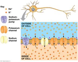
Ion Channels: Allow specific ions to cross membrane along electrochemical gradient.
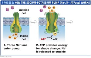
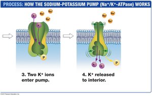
Na+/K+ ATPase: Maintains ion gradients by pumping Na+ out and K+ in.
Action Potential: Stages and Propagation
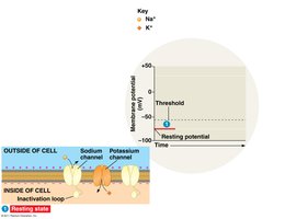
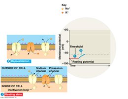
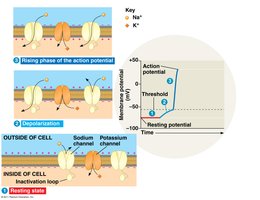
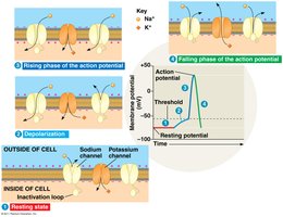
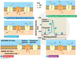
An action potential is a rapid change in membrane potential that propagates along the axon. It involves several stages: resting, depolarization, rising phase, falling phase, and undershoot.
Resting Potential: Most Na+ and K+ channels closed.
Depolarization: Na+ channels open, Na+ enters cell.
Rising Phase: Membrane potential increases.
Falling Phase: Na+ channels inactivate, K+ channels open, K+ exits cell.
Undershoot: K+ permeability high, then returns to resting via Na+/K+ ATPase.
Refractory Period: Na+ channels temporarily inactivated; prevents backward propagation.
Propagation and Myelination
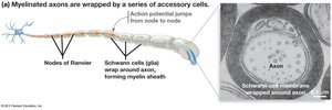
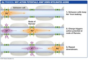
Action potentials propagate in one direction due to refractory Na+ channels. Axon diameter and myelination affect speed. Myelin sheaths, formed by glia (Schwann cells in PNS, oligodendrocytes in CNS), insulate axons and allow rapid signal transmission via saltatory conduction.
Large Diameter Axons: Faster transmission, less current leakage.
Myelin Sheath: Electrical insulation; action potentials jump between nodes of Ranvier.
Multiple Sclerosis: Degeneration of myelin impairs signaling.
Synaptic Transmission and Integration
Neurotransmitter Release and Synaptic Function
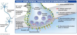
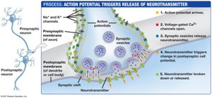
Action potentials trigger neurotransmitter release at synapses. Calcium ions enter the presynaptic cell, causing synaptic vesicles to fuse with the membrane and release neurotransmitters into the synaptic cleft.
Neurotransmitter Binding: Binds to ligand-gated ion channels on postsynaptic membrane.
Signal Transduction: Chemical signal converted to electrical signal.
Termination: Neurotransmitter broken down or reabsorbed.
Excitatory and Inhibitory Synapses (EPSPs and IPSPs)
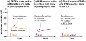
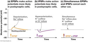
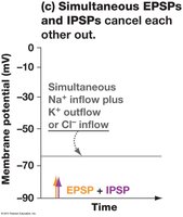
Synapses can be excitatory (EPSPs) or inhibitory (IPSPs). EPSPs depolarize the membrane, increasing action potential likelihood, while IPSPs hyperpolarize, decreasing likelihood. EPSPs and IPSPs are graded and additive, allowing complex integration of signals.
EPSP: Depolarization, Na+ influx.
IPSP: Hyperpolarization, K+ or Cl- influx.
Summation: Multiple EPSPs/IPSPs can combine; spatial and temporal summation.
Peripheral and Central Nervous System Functions
PNS: Afferent and Efferent Divisions
The peripheral nervous system (PNS) is divided into afferent (sensory) and efferent (motor) divisions. The afferent division transmits sensory information to the CNS, while the efferent division carries commands from the CNS to effectors.
Afferent Division: Monitors internal and external conditions.
Efferent Division: Controls body responses via motor neurons.
Efferent Division: Somatic and Autonomic Systems
The efferent division consists of the somatic system (voluntary control) and the autonomic system (involuntary control). The autonomic system is further divided into parasympathetic (relaxation/digestion) and sympathetic (fight or flight) systems.
Somatic System: Controls skeletal muscles; conscious control.
Autonomic System: Controls internal organs/glands; unconscious control.
Parasympathetic: Promotes relaxation and self-maintenance.
Sympathetic: Prepares for stress and arousal.
Major Structures of the Brain
The brain is composed of four major structures: cerebrum, cerebellum, diencephalon, and brain stem. The corpus callosum connects the two hemispheres.
Cerebrum: Conscious thought and memory; divided into hemispheres.
Cerebellum: Coordinates motor patterns.
Diencephalon: Relays sensory information; controls homeostasis.
Brain Stem: Autonomic center; connects brain to spinal cord.
Corpus Callosum: Thick band of axons connecting hemispheres.
Summary Table: Nervous System Components
Component | Function | Example |
|---|---|---|
CNS | Integrates sensory input, coordinates response | Brain, spinal cord |
PNS | Transmits signals between CNS and effectors | Sensory and motor neurons |
Neuron | Signal transmission | Sensory, motor, interneuron |
Glia | Support and insulate neurons | Schwann cells, oligodendrocytes |
Synapse | Signal transfer between cells | Neurotransmitter release |
Myelin Sheath | Insulation, rapid signal transmission | Schwann cell, node of Ranvier |
