 Back
BackNeurulation and Neuronal Signaling: From Morphogenesis to Action Potentials
Study Guide - Smart Notes
 Tailored notes based on your materials, expanded with key definitions, examples, and context.
Morphogenesis and Organogenesis
Neurulation: Formation of the Central Nervous System
Neurulation is a critical process in vertebrate development, leading to the formation of the brain and spinal cord. This process involves the transformation of the embryonic ectoderm into the neural plate, which then folds to form the neural tube—the precursor to the central nervous system (CNS).
Notochord: A rod-like structure derived from mesoderm cells that secretes signaling molecules to induce neural plate formation in the overlying ectoderm.
Neural Plate: A thickened region of ectoderm that gives rise to the neural tube.
Neural Tube: Formed by the folding and fusion of the neural plate; develops into the brain and spinal cord.
Neural Crest Cells: Cells at the border of the neural plate that migrate to form diverse structures, including peripheral nerves.
Somites: Blocks of mesoderm that segment alongside the neural tube and give rise to vertebrae and skeletal muscle.
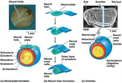
Example: Failure of the neural tube to close properly can result in neural tube defects such as spina bifida.
Neuronal Signaling
Neuron Structure and Function
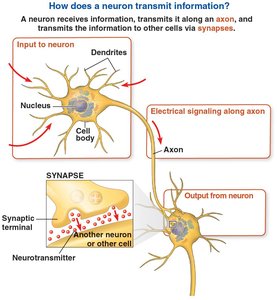
Neurons are the fundamental units of the nervous system, specialized for the reception, integration, and transmission of electrical and chemical signals.
Cell Body (Soma): Contains the nucleus and organelles; integrates incoming signals.
Dendrites: Branched extensions that receive signals from other neurons.
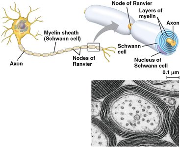
Axon: A single, long projection that transmits signals to other cells.
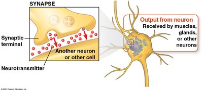
Synapse: The junction between a neuron and another cell where neurotransmitters mediate signal transmission.

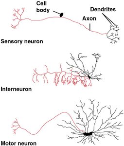
Types of Neurons
Sensory Neurons: Transmit information about external stimuli to the CNS.
Interneurons: Integrate information within the CNS.
Motor Neurons: Transmit signals from the CNS to muscles or glands.
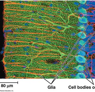
Supporting Cells: Glia
Glial Cells: Support, nourish, and protect neurons; maintain homeostasis and form myelin.
Organization of the Nervous System
Central Nervous System (CNS): Brain and spinal cord; responsible for integration and processing.
Peripheral Nervous System (PNS): Neurons outside the CNS; transmits information to and from the CNS.
Resting Potential
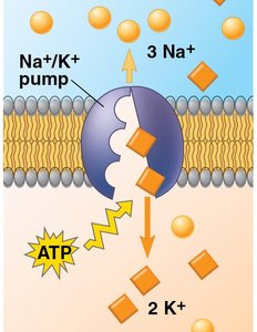
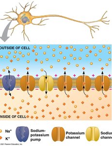
Establishment of Resting Membrane Potential
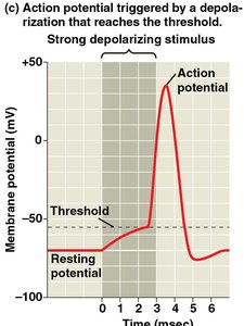
The resting potential is the voltage difference across the plasma membrane of a neuron at rest, typically between -60 and -80 mV. This potential is essential for the generation of action potentials.
Sodium-Potassium Pump: Uses ATP to actively transport 3 Na+ ions out and 2 K+ ions into the cell, maintaining ion gradients.
Ion Channels: Allow passive movement of ions; more open K+ channels than Na+ channels at rest, leading to a net negative charge inside the cell.
Action Potentials
Generation and Phases of Action Potentials
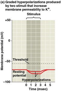
An action potential is a rapid, all-or-none change in membrane potential that propagates along the axon. It is triggered when depolarization reaches a threshold (about -55 mV).
Depolarization: Opening of voltage-gated Na+ channels causes Na+ influx.
Repolarization: Inactivation of Na+ channels and opening of K+ channels leads to K+ efflux.
Hyperpolarization: Membrane potential becomes more negative than resting potential due to continued K+ efflux.
Conduction of Action Potentials
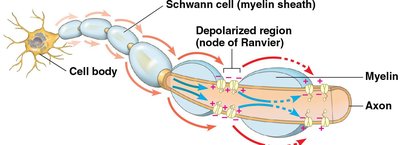
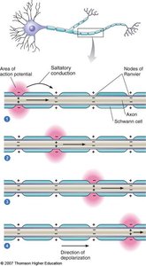
Action potentials travel unidirectionally along the axon. Their speed increases with axon diameter and the presence of myelin sheaths.
Myelin Sheath: Insulating layer produced by glia (Schwann cells in PNS, oligodendrocytes in CNS) that enables rapid signal conduction.
Nodes of Ranvier: Gaps in myelin where voltage-gated channels are concentrated; site of action potential regeneration.
Saltatory Conduction: Action potentials jump from node to node, increasing conduction velocity.
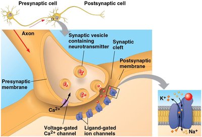
Synapses and Neurotransmitters
Chemical Synapses
At chemical synapses, neurotransmitters are released from the presynaptic neuron and bind to receptors on the postsynaptic cell, transmitting the signal across the synaptic cleft.
Synaptic Vesicles: Store neurotransmitters in the presynaptic terminal.
Neurotransmitter Release: Triggered by Ca2+ influx through voltage-gated channels.
Ligand-Gated Ion Channels: Open in response to neurotransmitter binding, altering postsynaptic membrane potential.
Major Neurotransmitters
Acetylcholine: Involved in muscle stimulation, memory, and learning.
Amino Acids: Glutamate (excitatory, memory), Glycine, GABA (inhibitory).
Biogenic Amines: Norepinephrine, epinephrine, dopamine, serotonin (mood, arousal, learning).
Neuropeptides: Substance P, endorphins (pain perception, mood).
Gases: Nitric oxide (NO), acts at inhibitory synapses.
Summary Table: Key Features of Neuronal Signaling
Feature | Description | Example |
|---|---|---|
Resting Potential | Voltage difference across membrane at rest (-60 to -80 mV) | Maintained by Na+/K+ pump |
Action Potential | Rapid depolarization and repolarization of membrane | Nerve impulse transmission |
Myelin Sheath | Insulating layer for fast conduction | Schwann cells in PNS |
Synapse | Junction for signal transmission between cells | Neuromuscular junction |
Neurotransmitter | Chemical messenger | Acetylcholine, GABA |
Additional info: The notes above integrate foundational concepts from chapters on animal development, nervous system structure, and neuronal signaling, providing a comprehensive overview suitable for college-level biology students.
