 Back
Osmoregulation and Excretion in Animals
Study Guide - Smart Notes
 Tailored notes based on your materials, expanded with key definitions, examples, and context.
Osmoregulation and Excretion
Overview of Osmoregulation and Excretion
Osmoregulation is the process by which organisms regulate the balance of water and solutes within their bodies, ensuring cellular function and overall homeostasis. Excretion refers to the removal of metabolic wastes, particularly nitrogenous wastes, from the body. In terrestrial organisms, these processes are closely linked due to the need to conserve water while eliminating toxic byproducts.
Osmoregulation: Maintains the concentration of solutes and water in body fluids.
Excretion: Eliminates metabolic wastes, especially nitrogenous compounds.
Key Organs: In mammals, the kidneys are the primary organs for both osmoregulation and excretion.
Homeostasis: The endocrine system (hormones) plays a critical role in maintaining fluid and solute balance.
Principles of Osmoregulation
Osmosis and Osmolarity
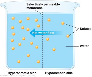
Osmosis is the movement of water across a selectively permeable membrane from regions of low solute concentration (high water potential) to regions of high solute concentration (low water potential). Osmolarity is a measure of solute concentration, expressed in milliosmoles per liter (mOsm/L).
Osmosis: Drives water movement in and out of cells and tissues.
Osmolarity: Blood (~300 mOsm/L), freshwater (~0-20 mOsm/L), seawater (~1000 mOsm/L).
Hyperosmotic: Solution with higher solute concentration.
Hypoosmotic: Solution with lower solute concentration.
Osmoconformers vs. Osmoregulators
Animals employ different strategies to manage osmotic balance depending on their environment:
Osmoconformers: Body fluids are isoosmotic with the environment (e.g., most marine invertebrates).
Osmoregulators: Actively regulate internal osmolarity, regardless of external conditions (e.g., vertebrates, freshwater and terrestrial animals).
Stenohaline: Tolerate only a narrow range of external osmolarities.
Euryhaline: Can survive large fluctuations in external osmolarity.
Osmoregulation in Vertebrates
Marine, Freshwater, and Terrestrial Vertebrates
Vertebrates have evolved distinct osmoregulatory strategies based on their habitats:
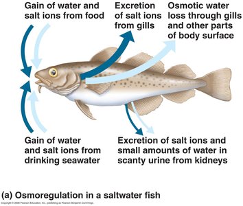
Marine Fish: Body fluids are hypoosmotic to seawater; lose water by osmosis and must drink seawater, excreting excess ions via gills and kidneys.
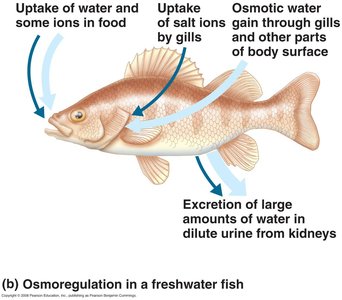
Freshwater Fish: Body fluids are hyperosmotic to freshwater; gain water by osmosis and excrete large volumes of dilute urine, actively taking up ions.
Terrestrial Tetrapods: Main challenge is dehydration; adaptations include water-tight skin, efficient kidneys, and behavioral strategies to conserve water.
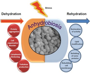
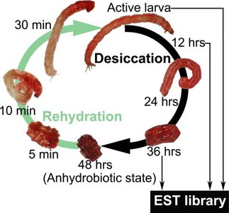
Anhydrobiosis
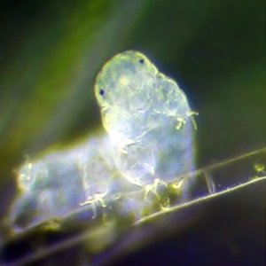
Some animals can survive extreme dehydration by entering a dormant state called anhydrobiosis, allowing them to lose almost all body water and later recover upon rehydration.
Examples: Tardigrades and some insect larvae.
Mechanisms: Production of protective molecules (e.g., trehalose), stabilization of proteins and membranes.
Nitrogenous Waste Products
Types of Nitrogenous Wastes
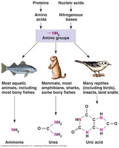
The metabolism of proteins and nucleic acids produces ammonia (NH3), which is toxic and must be excreted or converted to less toxic forms. The type of nitrogenous waste produced depends on the organism's habitat and evolutionary history.
Ammonia (NH3): Highly toxic, highly soluble; excreted directly by aquatic animals.
Urea: Less toxic, requires energy to produce; excreted by mammals, most amphibians, and some fish.
Uric Acid: Least toxic, insoluble, excreted as a paste or solid; produced by reptiles (including birds), insects, and land snails.
Trade-off: Energy cost vs. toxicity and water conservation.
Excretory Systems and the Mammalian Kidney
General Structure of Excretory Systems
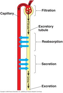
Most animal excretory systems share a basic design, involving filtration, reabsorption, secretion, and excretion. These processes allow the selective removal of wastes while retaining valuable solutes and water.
Filtration: Body fluid is forced through a selectively permeable membrane; water and small solutes pass, large molecules are retained.
Reabsorption: Valuable substances are reclaimed from the filtrate.
Secretion: Additional wastes are actively transported into the filtrate.
Excretion: The final filtrate (urine) is expelled from the body.
Structure and Function of the Mammalian Kidney
The mammalian kidney is a complex organ composed of an outer cortex and inner medulla, containing millions of nephrons—the functional units responsible for urine formation.
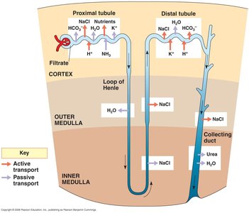
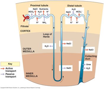
Nephron: Consists of Bowman's capsule, proximal tubule, loop of Henle, distal tubule, and collecting duct.
Blood Supply: Each nephron is closely associated with capillaries for exchange of substances.
Pathway: Filtrate flows from Bowman's capsule → proximal tubule → loop of Henle → distal tubule → collecting duct → ureter → bladder → urethra.
Osmolarity Gradient and Selective Reabsorption
Kidney function relies on an osmolarity gradient in the medulla and selective reabsorption of water and solutes at different nephron segments.
Proximal Tubule: Active reabsorption of ions and water; wastes remain in filtrate.
Descending Loop of Henle: Permeable to water, not salts; water is reabsorbed, concentrating the filtrate.
Ascending Loop of Henle: Permeable to NaCl, not water; salts are reabsorbed, diluting the filtrate.
Distal Tubule: Regulates NaCl and K+ exchange.
Collecting Duct: Final concentration of urine; permeability to water and urea is hormonally regulated.
Hormonal Regulation of Kidney Function
Antidiuretic Hormone (ADH)
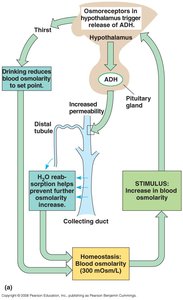
ADH is released in response to increased blood osmolarity. It acts on the distal tubules and collecting ducts to increase water reabsorption, concentrating urine and restoring osmotic balance.
Source: Produced in the hypothalamus, stored and released by the pituitary gland.
Effect: Increases permeability of collecting ducts to water.
Feedback: Drinking water and increased reabsorption lower blood osmolarity, reducing ADH release.
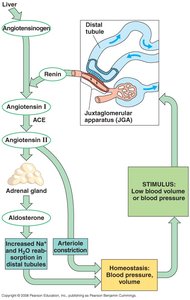
Renin-Angiotensin-Aldosterone System (RAAS)
The RAAS is activated when blood pressure or volume is low. It triggers a cascade of hormonal signals that increase sodium and water reabsorption, constrict blood vessels, and restore blood pressure and volume.
Stimulus: Low blood pressure or volume.
Effect: Increased Na+ and water reabsorption, arteriole constriction.
Outcome: Restores blood pressure and volume to homeostatic levels.
Summary Table: Nitrogenous Waste Types
Type of Waste | Example Organisms | Toxicity | Water Requirement | Energy Cost |
|---|---|---|---|---|
Ammonia | Most aquatic animals | High | High | Low |
Urea | Mammals, amphibians, sharks | Moderate | Moderate | Moderate |
Uric Acid | Reptiles, birds, insects | Low | Low | High |
