 Back
BackOsmoregulation and Excretion in Animals: Study Guide
Study Guide - Smart Notes
 Tailored notes based on your materials, expanded with key definitions, examples, and context.
Osmoregulation and Excretion in Animals
Introduction to Osmoregulation and Excretion
Animals must regulate the balance of water and solutes to survive in diverse environments. Osmoregulation is the process by which organisms control solute concentrations and balance water gain and loss. Excretion is the removal of metabolic waste products, especially nitrogenous wastes, from the body.
Osmoregulation: Balancing Water and Solutes
Osmosis and Osmolarity
Water moves across cell membranes by osmosis, driven by differences in osmolarity (solute concentration). The direction of water movement depends on the relative osmolarity of solutions separated by a selectively permeable membrane:
Isoosmotic solutions: No net movement of water; water crosses the membrane at equal rates in both directions.
Hypoosmotic to hyperosmotic: Water moves from the less concentrated (hypoosmotic) to the more concentrated (hyperosmotic) solution.
Osmoregulatory Strategies
Osmoconformers: Animals that are isoosmotic with their environment and do not actively regulate osmolarity (e.g., most marine invertebrates).
Osmoregulators: Animals that expend energy to control water uptake and loss in hyperosmotic or hypoosmotic environments, allowing them to inhabit a wider range of habitats.
Stenohaline vs. Euryhaline Animals
Stenohaline: Can tolerate only narrow changes in external osmolarity.
Euryhaline: Can survive large fluctuations in external osmolarity (e.g., salmon).
Osmoregulation in Different Environments
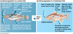
Marine Animals: Most marine invertebrates are osmoconformers, but marine bony fishes are hypoosmotic to seawater and must drink seawater, excreting excess salts through gills and kidneys.
Freshwater Animals: Constantly gain water by osmosis and lose salts by diffusion; they excrete large amounts of dilute urine and actively uptake salts through gills and food.
Terrestrial Animals: Adaptations such as waterproof body coverings, nocturnal behavior, and metabolic water production help reduce water loss.
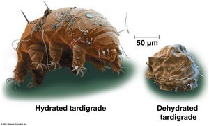
Animals in Temporary Waters: Some, like tardigrades, survive extreme dehydration through anhydrobiosis.
Energetics of Osmoregulation
Osmoregulation requires energy, with the cost depending on the difference between the animal's internal osmolarity and its environment, the permeability of surfaces, and the work required to transport solutes.
Transport Epithelia
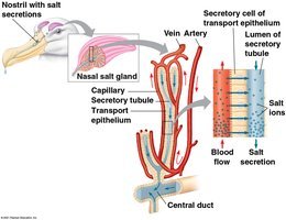
Transport epithelia are specialized cells that move solutes in specific directions, often forming tubular networks. An example is the nasal salt gland in marine birds, which removes excess sodium chloride from the blood.
Nitrogenous Wastes: Forms and Evolution
Types of Nitrogenous Waste
Ammonia: Highly toxic, requires large amounts of water for excretion; common in aquatic animals.
Urea: Less toxic, more energetically expensive to produce, requires less water; common in mammals and some marine species.
Uric Acid: Least toxic, excreted as a paste with minimal water loss; common in insects, land snails, reptiles, and birds.
The form of nitrogenous waste is influenced by phylogeny, habitat, water availability, and diet.
Excretory Systems: Structure and Function
General Functions of Excretory Systems
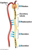
Most excretory systems produce urine by refining a filtrate derived from body fluids. The main steps are:
Filtration: Filtering of body fluids.
Reabsorption: Recovery of valuable solutes.
Secretion: Addition of nonessential solutes and wastes to the filtrate.
Excretion: Release of processed filtrate (urine) from the body.
Types of Excretory Systems
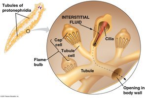
Protonephridia: Networks of dead-end tubules capped by flame bulbs; function in osmoregulation in flatworms.
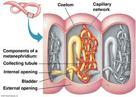
Metanephridia: Tubules in each segment of annelids (e.g., earthworms); function in both excretion and osmoregulation.
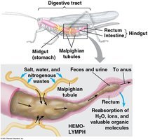
Malpighian Tubules: In insects and terrestrial arthropods; remove nitrogenous wastes from hemolymph and conserve water efficiently.
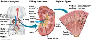
Kidneys: Excretory organs in vertebrates; highly organized tubules filter blood and regulate water and solute balance.
Nephron Structure and Function
Nephron Anatomy
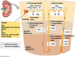
Each nephron consists of a long tubule and a ball of capillaries called the glomerulus, surrounded by the Bowman's capsule. The nephron processes filtrate through several regions:
Proximal tubule
Loop of Henle (descending and ascending limbs)
Distal tubule
Collecting duct
Steps of Filtrate Processing
Proximal Tubule: Reabsorption of ions, water, and nutrients; secretion of toxins.
Descending Limb of Loop of Henle: Water reabsorption via aquaporins; filtrate becomes concentrated.
Ascending Limb of Loop of Henle: Salt diffuses out; filtrate becomes dilute.
Distal Tubule: Regulates K+ and NaCl; pH regulation.
Collecting Duct: Final reabsorption of water and solutes; urine becomes hyperosmotic.
Solute Gradients and Water Conservation
Countercurrent Multiplier System
The countercurrent multiplier system in the loop of Henle maintains a high salt concentration in the kidney, allowing for the production of hyperosmotic urine. This adaptation is crucial for water conservation in terrestrial animals.
Adaptations in Vertebrate Kidneys
Mammals: Long loops of Henle in desert species for water conservation; short loops in aquatic species.
Birds and Reptiles: Birds have shorter loops; excrete uric acid. Reptiles reabsorb water in the cloaca and excrete uric acid.
Freshwater Fishes and Amphibians: Excrete dilute urine; reabsorb ions in distal tubules.
Marine Bony Fishes: Fewer, smaller nephrons; rely on gill chloride cells for osmoregulation.
Hormonal Regulation of Kidney Function
Antidiuretic Hormone (ADH)
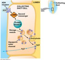
ADH (vasopressin) is released from the posterior pituitary and increases water reabsorption in the collecting ducts by promoting the insertion of aquaporin channels. This reduces urine volume and concentrates urine.
Osmoreceptors and Feedback Control
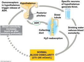
Osmoreceptors in the hypothalamus monitor blood osmolarity and regulate ADH release. Increased osmolarity triggers ADH release, leading to water conservation; decreased osmolarity suppresses ADH, resulting in dilute urine.
Renin-Angiotensin-Aldosterone System (RAAS)
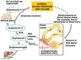
The RAAS is activated by a drop in blood pressure near the glomerulus, leading to the release of renin, formation of angiotensin II, vasoconstriction, and aldosterone release. This increases sodium and water reabsorption, raising blood pressure and volume.
Atrial Natriuretic Peptide (ANP)
ANP is released in response to increased blood volume and pressure. It inhibits renin release, reducing blood pressure and volume, and opposes the actions of RAAS and ADH.
Summary Table: Comparison of Nitrogenous Wastes
Waste Type | Toxicity | Water Requirement | Energy Cost | Main Excretors |
|---|---|---|---|---|
Ammonia | High | High | Low | Aquatic animals |
Urea | Moderate | Moderate | Moderate | Mammals, amphibians, some fish |
Uric Acid | Low | Low | High | Insects, birds, reptiles |
Key Equations
Osmosis: Water moves from regions of low solute concentration to high solute concentration across a selectively permeable membrane.
Osmolarity (Osm):
