 Back
BackOsmoregulation and Excretion: Mechanisms and Structures in Animals
Study Guide - Smart Notes
 Tailored notes based on your materials, expanded with key definitions, examples, and context.
Osmoregulation and Excretion
Introduction to Osmoregulation and Excretion
Osmoregulation and excretion are essential physiological processes that maintain homeostasis in animals by regulating water and solute balance and removing metabolic wastes. These processes are critical for cellular function and overall organismal health.

Functions of the Urinary (Excretory) System
Main Functions
Osmoregulation: Maintains a constant fluid-solute balance in the body, ensuring that extracellular ions (Na+, Cl-, K+, Ca++, Mg++) remain within optimal ranges for cellular function.
Excretion of Nitrogenous Wastes: Removes toxic byproducts of protein and nucleic acid metabolism, such as ammonia, urea, and uric acid.
Osmoregulation
Osmoregulation is a type of homeostatic balance that maintains the proper concentration of solutes and water in the body. The unit for solute concentration is osmolarity, measured in milliosmoles per liter (mOsm/L).
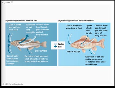
Freshwater organisms: Hyperosmotic to their environment; water enters their bodies, so they excrete large amounts of dilute urine to prevent bursting.
Saltwater organisms: Hypoosmotic to their environment; water leaves their bodies, so they drink seawater and excrete concentrated urine to prevent dehydration.
Osmoconformers: Isoosmotic with their environment; do not need to actively regulate water balance.
Terrestrial organisms: Lose water through evaporation and excretion; adaptations include waxy cuticles, exoskeletons, and keratinized skin.
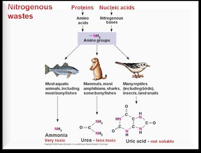
Excretion of Nitrogenous Wastes
Types of Nitrogenous Wastes
Ammonia (NH3): Highly toxic, excreted by aquatic animals (e.g., fish); requires large amounts of water for dilution.
Urea: Less toxic, excreted by mammals and amphibians; soluble in water and requires energy (ATP) to synthesize from ammonia.
Uric Acid: Nontoxic, excreted by birds and reptiles; insoluble in water, excreted as a semisolid paste, energetically expensive to produce.
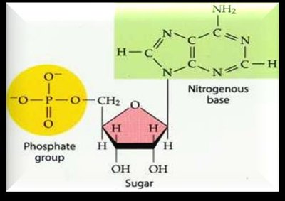
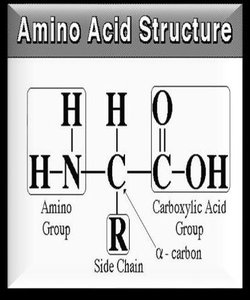
Nitrogenous wastes are produced from the metabolism of proteins and nucleic acids, which contain amino groups.
Mechanisms of Filtration in Excretory Systems
Overview of Filtration Mechanisms
Contractile Vacuole (Protozoans)
Flame Cells in Protonephridia (Flatworms)
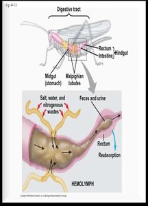
Malpighian Tubules (Insects)
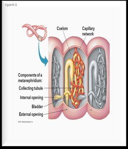
Nephridia (Annelids)
Kidneys (Vertebrates)
Contractile Vacuole
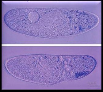
Found in freshwater protozoans like Paramecium, the contractile vacuole collects excess water and expels it to maintain osmotic balance.
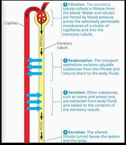
General Steps in Excretory Filtration
Filtration: Body fluids are forced through a selectively permeable membrane by hydrostatic pressure; small molecules pass, large molecules are retained.
Reabsorption: Useful substances are actively transported back into the body fluids.
Secretion: Additional wastes are actively transported into the filtrate.
Excretion: The final filtrate (urine) is expelled from the body.
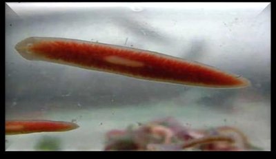
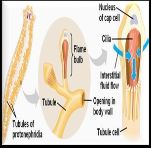
Flame Cells in Protonephridia
Found in flatworms (e.g., planaria), protonephridia are networks of tubules with flame cells that filter and expel wastes. Cilia in flame cells drive fluid movement.
Malpighian Tubules
In insects, Malpighian tubules extend from the gut and actively transport wastes and ions into the tubules, with water following by osmosis. Wastes are excreted with feces.
Nephridia (Metanephridia)
In annelids (e.g., earthworms), metanephridia are segmentally arranged excretory organs that filter coelomic fluid, reabsorb useful substances, and excrete wastes through external pores.
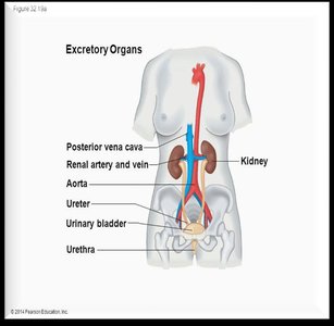
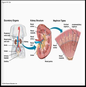
Human Excretory System
Major Organs and Structures
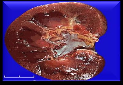
Kidneys: Filter blood and produce urine; located in the retroperitoneal cavity.
Ureters: Transport urine from kidneys to bladder.
Bladder: Stores urine until excretion.
Urethra: Conducts urine out of the body.
Other Functions of the Kidney
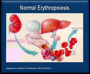
Hormone Production: Renin (regulates blood pressure) and erythropoietin (stimulates red blood cell production, especially at high altitudes).
Vitamin D Activation: Converts vitamin D to its active form for calcium absorption.
Kidney Structure and Nephron Organization
Kidney Anatomy
Renal Cortex: Outer region, contains most nephrons.
Renal Medulla: Inner region, contains loops of Henle and collecting ducts.
Renal Pelvis: Central cavity where urine collects before entering the ureter.
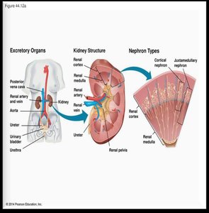
Nephron: The Functional Unit
Each kidney contains about 1 million nephrons.
Cortical nephrons: Located in the cortex (85% of nephrons).
Juxtamedullary nephrons: Extend into the medulla; crucial for urine concentration.
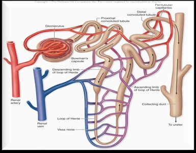
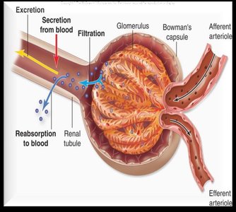
Glomerulus: Ball of capillaries where filtration begins.
Bowman's Capsule: Surrounds the glomerulus and collects filtrate.

Order of Filtrate Flow in the Nephron
Bowman's Capsule → Proximal Tubule → Loop of Henle (Descending and Ascending limbs) → Distal Tubule → Collecting Duct
From the collecting duct: Renal Pelvis → Ureters → Bladder → Urethra
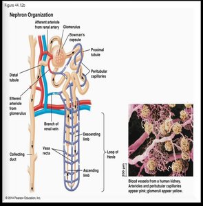
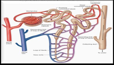
Capillary System in the Kidney
Afferent arteriole: Brings blood to the glomerulus.
Efferent arteriole: Carries blood away from the glomerulus.
Peritubular capillaries: Surround proximal and distal tubules for reabsorption and secretion.
Vasa recta: Capillaries that serve the juxtamedullary nephrons and loop of Henle.

Processes of Excretion in the Nephron
Filtration
Filtration occurs at the glomerulus and Bowman's capsule, driven by high hydrostatic pressure (about 55 mm Hg). Small molecules (water, urea, ions, glucose, amino acids) pass into the filtrate; large proteins and cells remain in the blood.
Proximal Tubule
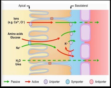
The proximal tubule reabsorbs about 99% of water, amino acids, glucose, ions, and bicarbonate back into the blood. It is lined with simple cuboidal epithelial cells with microvilli to increase surface area for absorption.
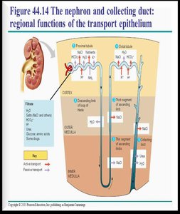
Loop of Henle
Descending limb: Permeable to water but not salts; water exits by osmosis, concentrating the filtrate.
Ascending limb: Impermeable to water; Na+ and Cl- are reabsorbed (passively in the thin segment, actively in the thick segment), diluting the filtrate.
The loop of Henle establishes a countercurrent multiplier system, crucial for concentrating urine in terrestrial vertebrates.
Distal Tubule and Collecting Duct
Distal Tubule: Involved in secretion (HCO3-, NaCl, water) and regulated by hormones such as aldosterone.
Collecting Duct: Final site for water reabsorption, regulated by antidiuretic hormone (ADH); urine becomes highly concentrated as it passes through the hyperosmotic medulla.
Summary Table: Main Steps and Structures in Excretion
Step | Location | Main Function |
|---|---|---|
Filtration | Glomerulus/Bowman's Capsule | Initial removal of small molecules from blood |
Reabsorption | Proximal Tubule, Loop of Henle, Distal Tubule, Collecting Duct | Return of useful substances to blood |
Secretion | Distal Tubule, Collecting Duct | Active removal of additional wastes from blood |
Excretion | Collecting Duct → Ureter → Bladder → Urethra | Removal of urine from body |
Hormonal Regulation of Kidney Function
Key Hormones
Aldosterone: Secreted by the adrenal cortex; increases sodium reabsorption in the distal tubule, leading to water retention and increased blood pressure.
Antidiuretic Hormone (ADH, Vasopressin): Secreted by the posterior pituitary; increases water reabsorption in the collecting duct by increasing aquaporin channels, concentrating urine and conserving water.
Diuretics: Substances that promote urine production by inhibiting ADH (e.g., alcohol, caffeine, some medications).
Histology of the Nephron
Cell Types
Simple Cuboidal Epithelium: Lines most of the nephron tubule, specialized for absorption and secretion.
Microvilli: Increase surface area for reabsorption in the proximal tubule.
Clinical Connections (Not on Exam)
Kidney Failure
Acute: Treatable; may require temporary dialysis.
Chronic: Irreversible; requires transplant or long-term dialysis.
Kidney Stones
Formed from high concentrations of minerals (e.g., calcium, uric acid) in urine.
Can cause pain and block urinary flow.
Additional info: The nephron's countercurrent multiplier system is analogous to countercurrent exchange in fish gills, maximizing efficiency of solute and water movement.
