 Back
BackPeripheral Nervous System: Structure, Function, and Divisions
Study Guide - Smart Notes
 Tailored notes based on your materials, expanded with key definitions, examples, and context.
Peripheral Nervous System (PNS)
Overview of the Nervous System
The nervous system is divided into two main parts: the Central Nervous System (CNS) and the Peripheral Nervous System (PNS). The CNS consists of the brain and spinal cord, while the PNS comprises nerves and ganglia that connect the CNS to the rest of the body, including receptors, muscles, and glands.
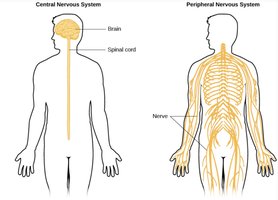
CNS: Brain and spinal cord; responsible for processing and integrating information.
PNS: Nerves and ganglia outside the CNS; transmits signals between the CNS and the body.
Peripheral Nerves
The PNS includes cranial nerves (12 pairs arising from the brain) and spinal nerves (31 pairs arising from the spinal cord). These nerves are essential for communication between the CNS and peripheral tissues.
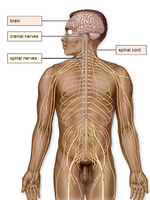
Cranial nerves: Serve the head and neck region; some are sensory, some motor, most are mixed.
Spinal nerves: All are mixed nerves, containing both sensory and motor fibers.
Structure of Peripheral Nerves
Sensory, Motor, and Mixed Nerves
Nerves in the PNS can be classified based on the direction of impulse transmission:
Sensory (Afferent) fibers: Carry impulses into the CNS from receptors.
Motor (Efferent) fibers: Carry impulses away from the CNS to effectors (muscles/glands).
Mixed nerves: Contain both sensory and motor fibers.
Spinal Nerve Roots
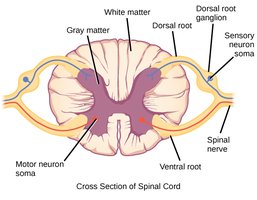
Each spinal nerve is attached to the spinal cord by two roots:
Ventral root: Contains axons of motor neurons; cell bodies are in the spinal cord's gray matter.
Dorsal root: Contains axons of sensory neurons; cell bodies are in the dorsal root ganglion.
Divisions of the Peripheral Nervous System
Functional Organization
The PNS is functionally divided into afferent (sensory) and efferent (motor) divisions:

Afferent (Sensory) Division: Transmits sensory information to the CNS.
Somatic sensory neurons: From skin, muscles, and joints.
Visceral sensory neurons: From internal organs.
Efferent (Motor) Division: Transmits motor commands from the CNS to effectors.
Somatic division: Controls voluntary movements of skeletal muscles.
Autonomic division: Controls involuntary actions of smooth muscle, cardiac muscle, and glands.
Autonomic Nervous System (ANS)
The ANS regulates involuntary physiological functions to maintain homeostasis. It operates largely without conscious control and is regulated by the medulla oblongata, hypothalamus, and cerebral cortex.
Sympathetic division: Prepares the body for 'fight-or-flight' responses; releases noradrenaline.
Parasympathetic division: Maintains 'rest-and-digest' functions; releases acetylcholine.

Neural Pathways in the PNS
Somatic vs. Autonomic Pathways
Somatic pathway: One motor neuron from CNS to skeletal muscle; neurotransmitter is acetylcholine.
Autonomic pathway: Two motor neurons (preganglionic and postganglionic); neurotransmitters are acetylcholine or noradrenaline.


Comparison of Autonomic and Somatic Divisions
Key Differences
Characteristic | Autonomic Division | Somatic Division |
|---|---|---|
Effectors | Heart muscle, involuntary muscles, glands | Skeletal (voluntary) muscles |
General Function | Adjusts internal environment | Response to external environment |
Efferent (outward) pathway | Two neurons from CNS to effector with a synapse in a ganglion | One neuron from CNS to effector |
Neurotransmitter at effector | Acetylcholine or noradrenaline | Acetylcholine |
Control | Usually involuntary | Usually voluntary |
Nerves to target organ | Two sets: sympathetic and parasympathetic | One set |
Effect on target organ | Excitation or inhibition | Always excitation |

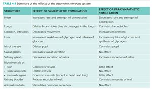
Effects of the Autonomic Nervous System
Sympathetic vs. Parasympathetic Stimulation
Structure | Effect of Sympathetic Stimulation | Effect of Parasympathetic Stimulation |
|---|---|---|
Heart | Increases rate and strength of contraction | Decreases rate and strength of contraction |
Lungs | Dilates bronchioles | Constricts bronchioles |
Liver | Increases breakdown of glycogen and release of glucose | Increases uptake of glucose and synthesis of glycogen |
Eye (pupil) | Dilates pupil | Constricts pupil |
Sweat glands | Increases secretion | No effect |
Blood vessels (skeletal muscle) | Dilates vessels | Little effect |
Adrenal medulla | Stimulates hormone secretion | No effect |
Fight-or-Flight Response
Sympathetic Activation
The fight-or-flight response is triggered by the sympathetic division during situations of fear, anger, stress, or danger. This response prepares the body for increased physical activity by:
Increasing heart rate and blood pressure
Dilating airways and blood vessels in muscles
Raising blood glucose levels
Stimulating sweat gland secretion
Releasing adrenaline and noradrenaline from the adrenal medulla
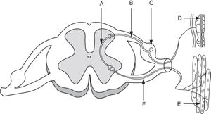
Neural Circuitry: Reflex Arc
Identification of Neurons
In a typical reflex arc, the afferent neuron (sensory neuron) transmits impulses to the CNS, the interneuron processes the information, and the efferent neuron (motor neuron) carries the response to the effector.
Description | Marks |
|---|---|
Afferent neuron = B | 1 |
Interneuron = A | 1 |
Total | 2 |
Additional info: The PNS is essential for integrating sensory input and motor output, and its divisions allow for both voluntary and involuntary control of body functions. Understanding the structure and function of the PNS is fundamental for studies in neurobiology, physiology, and medicine.
