 Back
BackPlant Cells, Tissues, and Organs: Structure and Function
Study Guide - Smart Notes
 Tailored notes based on your materials, expanded with key definitions, examples, and context.
Plant Cell Structure and Cell Theory
Cell Theory and Organism Theory
Plant biology is grounded in the principles of cell theory, which states that all living organisms are composed of one or more cells, all cells arise from preexisting cells, and the cell is the basic structural and functional unit of life. Organism theory, in contrast, emphasizes the organism as a whole as the functional unit, with plant cells often forming a continuous mass of protoplasm rather than acting as independent units.
Cell Theory: Foundation of modern biology; highlights the universality and continuity of cellular life.
Organism Theory: Recognizes the integrated nature of plant tissues, where cells are interconnected and not completely separate.
Plant Cell Wall
Structure and Function
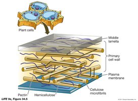
The plant cell wall is an extracellular structure that provides support, maintains internal pressure, and protects against pathogens. It is located outside the plasma membrane and is a defining feature of plant cells.
Primary Cell Wall: Composed of cellulose microfibrils embedded in a matrix of hemicellulose and pectin. It is the first wall formed, present in all plant cells, and is relatively soft and flexible, allowing for growth and permeability.
Middle Lamella: A pectin-rich layer that cements adjacent plant cells together.
Secondary Cell Wall: Formed inside the primary wall in some cells, it is thicker, more rigid, and often lignified, providing additional strength and waterproofing. Not all plant cells develop a secondary wall.
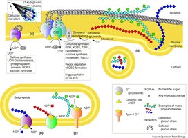
Cell Wall Composition and Synthesis
The synthesis of the cell wall involves complex biochemical pathways, with cellulose synthase complexes producing cellulose microfibrils at the plasma membrane, and other enzymes synthesizing hemicellulose and pectin.
Cellulose: The main structural component, forming strong microfibrils.
Hemicellulose and Pectin: Matrix polysaccharides that provide flexibility and adhesion.
Lignin: A complex hydrocarbon polymer added to secondary walls for rigidity and waterproofing.
Intercellular Connections
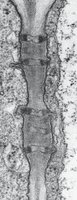
Plasmodesmata
Plasmodesmata are microscopic channels that traverse the cell walls, connecting the cytoplasm of adjacent plant cells. They allow for the movement of water, ions, and small molecules, facilitating communication and transport between cells.
Function: Enable cytoplasmic streaming and the passage of endoplasmic reticulum between cells.
Comparison: Functionally similar to gap junctions in animal cells.
Other Plant Cell Structures
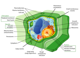
Vacuole and Chloroplast
Plant cells contain specialized organelles that are essential for their function and survival.
Vacuole: Occupies most of the cell volume; stores water, proteins, starch, and waste products. Also helps maintain turgor pressure.
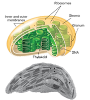
Chloroplast: Site of photosynthesis; contains chlorophyll within thylakoid membranes.
Plant Organs and Their Functions
The Three Main Plant Organs
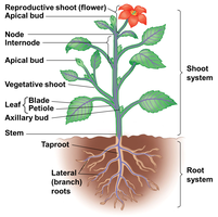
Plants are organized into three primary organs, each with specialized functions:
Stem: Provides structural support and transports water, minerals, and nutrients.
Leaf: Main site of photosynthesis.
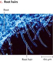
Root: Absorbs water and minerals from the soil.
Flowers and Fruits: Modified leaves and/or stems involved in reproduction.
Plant Tissues
Overview of Tissue Types
Plant tissues are classified into three main types based on their structure and function:
Dermal Tissue: Protective outer covering; includes the epidermis and cuticle.
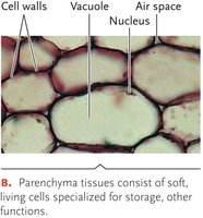
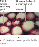
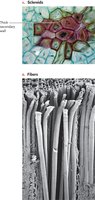
Ground Tissue: Fills the interior of the plant; involved in storage, support, and photosynthesis.
Vascular Tissue: Specialized for transport of water, minerals, and nutrients; includes xylem and phloem.
Dermal Tissue
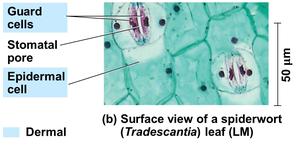
The epidermis forms the outermost layer of the primary plant body and is often covered by a waxy cuticle to minimize water loss. In leaves, pairs of guard cells in the epidermis form stomata, which regulate gas exchange.
Cuticle: Waxy layer that restricts water loss.
Stomata: Openings controlled by guard cells for gas exchange.
Ground Tissue Types
Ground tissue is divided into three main cell types, each with distinct roles:
Parenchyma: Soft, living cells with thin primary walls; involved in storage, secretion, and photosynthesis.
Collenchyma: Elongated cells with unevenly thickened primary walls; provide flexible support while allowing growth.
Sclerenchyma: Cells with thick, lignified secondary walls; provide rigid support and protection. Includes sclereids and fibers.
Vascular Tissue
Xylem
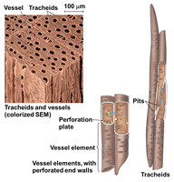
Xylem is responsible for conducting water and dissolved minerals from roots to leaves. It consists of dead cells with thick, lignified secondary walls.
Tracheids: Elongated, tapered cells with overlapping ends; water moves through pits.
Vessel Elements: Shorter, wider cells aligned end-to-end to form continuous tubes; have perforation plates for efficient water flow.
Phloem
Phloem transports the products of photosynthesis (mainly sugars) throughout the plant. Unlike xylem, phloem cells are alive at maturity.
Sieve Tube Members: Main conducting cells; lack a nucleus at maturity and are assisted by companion cells.
Companion Cells: Support sieve tube members metabolically and help load/unload sugars.
Organization of Tissues in Plant Organs
Leaves
Leaves are organized into dermal, ground, and vascular tissues. The upper and lower epidermis are covered by a cuticle, and the internal ground tissue is differentiated into palisade and spongy mesophyll for photosynthesis and gas exchange. Vascular bundles (veins) contain xylem and phloem.
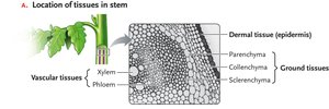
Stems
Vascular tissues in stems are organized into bundles, with phloem on the outside and xylem on the inside, often surrounded by sclerenchyma for support. The arrangement differs between monocots and eudicots.
Roots
Roots have a central vascular cylinder surrounded by ground tissue (cortex) and an outer epidermis. The endodermis regulates the flow of substances into the vascular cylinder, and the pericycle gives rise to lateral roots.
Organ | Dermal Tissue | Ground Tissue | Vascular Tissue |
|---|---|---|---|
Leaf | Epidermis, cuticle, guard cells | Palisade and spongy mesophyll | Xylem, phloem (veins) |
Stem | Epidermis, cuticle | Cortex, pith, collenchyma, sclerenchyma | Vascular bundles (xylem, phloem) |
Root | Epidermis, root hairs | Cortex, endodermis, pericycle | Vascular cylinder (xylem, phloem) |
Example: In a eudicot stem, vascular bundles are arranged in a ring, while in monocots, they are scattered throughout the ground tissue.
Additional info: The organization of tissues in plant organs is crucial for their specialized functions, such as efficient transport in stems, absorption in roots, and photosynthesis in leaves.
