 Back
BackProkaryotes and Microbial Diversity: Structure, Classification, and Cell Walls
Study Guide - Smart Notes
 Tailored notes based on your materials, expanded with key definitions, examples, and context.
Prokaryotes: Structure, Diversity, and Classification
Introduction to Microbiology and Prokaryotes
Microbiology is the study of microscopic organisms, including bacteria, archaea, fungi, viruses, and some algae and protozoa. Prokaryotes, which include bacteria and archaea, are among the most ancient and diverse forms of life on Earth. They are characterized by their simple cell structure, lack of a membrane-bound nucleus, and remarkable adaptability to extreme environments.
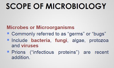
Microbes are commonly referred to as "germs" or "bugs" and include bacteria, fungi, algae, protozoa, and viruses.
Prokaryotes are unicellular organisms lacking a true nucleus and membrane-bound organelles.
They are found in nearly every environment, including extreme habitats such as salt lakes, thermal springs, and arctic regions.
Prokaryotes are divided into two domains: Bacteria and Archaea.
Size and Abundance of Microbes
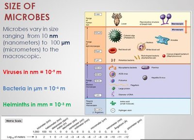
Microbes vary greatly in size, from nanometers to millimeters, and are the most abundant organisms on Earth. Prokaryotic cells are typically much smaller than eukaryotic cells.
1 nanometer (nm) = 0.000001 mm
1 micrometer (µm) = 0.001 mm
Most prokaryotic cells are 0.5–5 µm in diameter, while eukaryotic cells are 10–100 µm.
Viruses are even smaller, typically 10–100 nm.
Evolutionary History and Adaptation
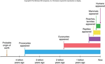
Prokaryotes are among the earliest life forms, with fossil evidence dating back 3.5 billion years. Their evolutionary success is attributed to their metabolic diversity and ability to adapt to a wide range of environments.
Prokaryotes appeared approximately 3.5 billion years ago, predating eukaryotes by over a billion years.
They are considered masters of adaptation due to their ability to thrive in diverse and extreme habitats.
Microbial Taxonomy and the Three-Domain System
Historically, prokaryotes were classified based on phenotypic traits such as shape, motility, and staining properties. Modern taxonomy uses molecular data, particularly ribosomal RNA sequences, to classify life into three domains: Bacteria, Archaea, and Eukarya.
The Woese-Fox Three Domain System is based on genetic relatedness, cell membrane lipid structure, and sensitivity to antibiotics.
Many prokaryotes once classified as bacteria are now recognized as archaea, which are more closely related to eukaryotes than to bacteria.
Comparative Cellular Structures of Prokaryotes and Eukaryotes
Prokaryotic cells differ fundamentally from eukaryotic cells in their structure and organization.
Prokaryotes lack a membrane-bound nucleus and organelles.
Eukaryotes possess a true nucleus and various membrane-bound organelles.

Key Differences Among Bacteria, Archaea, and Eukarya
The three domains of life can be distinguished by several cellular and molecular characteristics.
Characteristic | Bacteria | Archaea | Eukarya |
|---|---|---|---|
Nuclear envelope | Absent | Absent | Present |
Membrane-enclosed organelles | Absent | Absent | Present |
Peptidoglycan in cell wall | Present | Absent | Absent |
Membrane lipids | Unbranched hydrocarbons | Some branched hydrocarbons | Unbranched hydrocarbons |
RNA polymerase | One kind | Several kinds | Several kinds |
Initiator amino acid for protein synthesis | Formyl-methionine | Methionine | Methionine |
Introns in genes | Very rare | Present in some genes | Present in many genes |
Response to antibiotics | Growth inhibited | Growth not inhibited | Growth not inhibited |
Histones associated with DNA | Absent | Present in some species | Present |
Circular chromosome | Present | Present | Absent |
Growth at >100°C | No | Some species | No |
Characteristics of Bacteria and Archaea
Bacteria (Eubacteria): Unicellular, cell walls contain peptidoglycan, unbranched fatty acid chains, unique rRNA, some form endospores, both pathogenic and non-pathogenic species.
Archaea (Archaebacteria): Unicellular, no peptidoglycan, branched hydrocarbon chains, unique rRNA, often extremophiles (methanogens, halophiles, thermophiles).
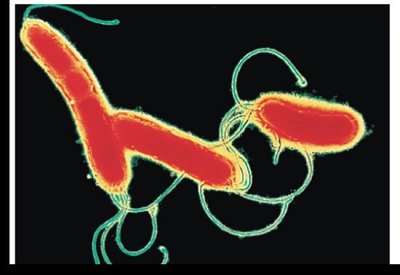
Prokaryotic Cell Morphology and Structure
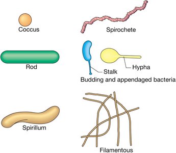
Common Shapes of Prokaryotic Cells
Prokaryotic cells exhibit a variety of shapes, which are important for identification and classification.
Cocci: Spherical; can occur singly (coccus), in pairs (diplococci), chains (streptococci), or clusters (staphylococci).
Bacilli: Rod-shaped; usually solitary (bacillus) or in chains (streptobacilli).
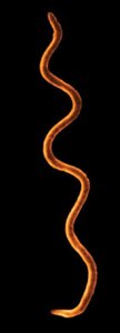
Spirilla/Spirillum: Spiral-shaped; range from comma-like to loose coils, including spirochetes.
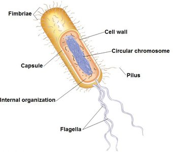
Typical Structures of a Prokaryotic Cell
Despite their simplicity, prokaryotic cells possess several key structures that enable survival and adaptation.
Cell wall: Provides shape, protection, and prevents lysis in hypotonic environments.
Plasma membrane: Regulates transport of substances in and out of the cell.
Nucleoid: Region containing the circular DNA chromosome.
Ribosomes: Sites of protein synthesis.
Flagella: Used for motility in some species.
Capsule: Protective outer layer found in some bacteria.
Pili/Fimbriae: Surface structures for attachment or conjugation.
Prokaryotic Cell Walls and Gram Staining
Composition and Function of Prokaryotic Cell Walls
The cell wall is a critical feature of prokaryotic cells, providing structural support and protection. The composition of the cell wall differs between bacteria and archaea.
Bacterial cell walls contain peptidoglycan (murein), a network of sugar polymers cross-linked by polypeptides.
Peptidoglycan is composed of N-acetylglucosamine (NAG) and N-acetylmuramic acid (NAM).
Archaeal cell walls lack peptidoglycan and may contain glycoproteins, polysaccharides, or pseudomurein.
Eukaryotic cell walls (when present) are made of cellulose (plants) or chitin (fungi).
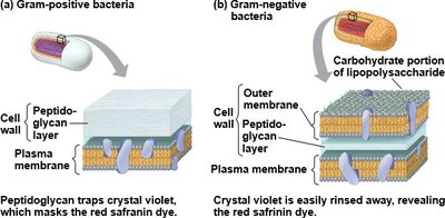
Gram Staining: Differentiating Bacterial Cell Walls
The Gram stain is a differential staining technique used to classify bacteria based on cell wall composition.
Gram-positive bacteria: Thick peptidoglycan layer, stain purple.
Gram-negative bacteria: Thin peptidoglycan layer, outer membrane with lipopolysaccharides (LPS), stain pink/red.
The lipid portion of LPS in Gram-negative bacteria is often toxic and provides resistance to certain antibiotics and host defenses.
Many antibiotics (e.g., penicillin) target peptidoglycan synthesis, making Gram-positive bacteria more susceptible.
Gram Staining Procedure
The Gram stain involves several steps to differentiate bacteria:
Application of crystal violet (primary stain)
Application of iodine (mordant)
Alcohol wash (decolorization)
Application of safranin (counterstain)
Gram-positive bacteria retain the crystal violet and appear purple, while Gram-negative bacteria lose the violet stain and take up the red safranin.
Summary Table: Gram-Positive vs. Gram-Negative Bacteria
Feature | Gram-Positive | Gram-Negative |
|---|---|---|
Peptidoglycan layer | Thick | Thin |
Outer membrane | Absent | Present (with LPS) |
Stain color | Purple | Pink/Red |
Antibiotic susceptibility | More susceptible | Less susceptible |
Toxicity | Usually less toxic | Lipid A of LPS is toxic |
Conclusion
Prokaryotes are fundamental to life on Earth, displaying remarkable diversity in structure, metabolism, and ecological roles. Understanding their classification, cell structure, and cell wall composition is essential for studying microbiology, evolution, and the impact of microbes on health and the environment.
