 Back
BackProkaryotes and Microbial Diversity: Structure, Classification, and Cell Walls
Study Guide - Smart Notes
 Tailored notes based on your materials, expanded with key definitions, examples, and context.
Introduction to Microbiology and Prokaryotes
Scope and Importance of Microbiology
Microbiology is the study of microscopic organisms, including bacteria, archaea, fungi, algae, protozoa, and viruses. These organisms, collectively called microbes, play essential roles in ecosystems, human health, and biotechnology.
Microbes are commonly referred to as “germs” or “bugs.”
They include bacteria, fungi, algae, protozoa, and viruses.
Prions (infectious proteins) are a recent addition to the list of microorganisms.
Why study microbiology? Microbes affect every aspect of human society and the natural world, influencing health, industry, and the environment.
Size and Abundance of Microbes
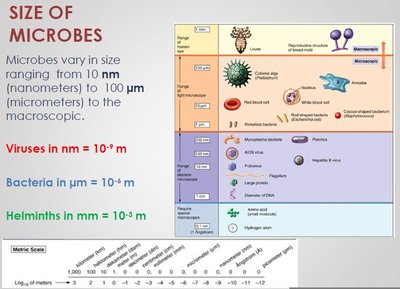
Microbes vary greatly in size, from nanometers (viruses) to millimeters (helminths). Bacteria are typically measured in micrometers (µm).
Viruses: 10-9 meters (nm)
Bacteria: 10-6 meters (µm)
Helminths: 10-3 meters (mm)
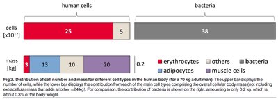
“Back of the envelope” calculations suggest that the average human contains more bacterial DNA than human DNA, highlighting the abundance and significance of microbes in and on our bodies.
Prokaryotes: Structure, Diversity, and Classification
Characteristics of Prokaryotes
Prokaryotes are microscopic, unicellular organisms lacking a membrane-bound nucleus and specialized organelles. They include bacteria and archaea and are among the most abundant and adaptable organisms on Earth.
Most prokaryotes are 0.5–5 µm in size, much smaller than most eukaryotic cells (10–100 µm).
They thrive in diverse and often extreme environments (e.g., salt lakes, thermal springs).
Prokaryotes are divided into two domains: Bacteria and Archaeabacteria.
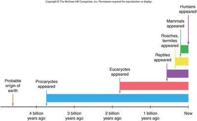
Evolutionary Timeline and Taxonomy
Bacteria appeared approximately 3.5 billion years ago, predating the evolution of eukaryotes by billions of years. Modern taxonomy uses molecular systematics, such as ribosomal RNA sequences, to classify prokaryotes into three domains: Bacteria, Archaea, and Eukarya.
Traditional classification relied on phenotypic traits (shape, motility, Gram staining).
Molecular data revealed that archaea are more closely related to eukaryotes than to bacteria.


Comparison of Bacteria, Archaea, and Eukarya
The three domains of life differ in several fundamental cellular characteristics, including cell wall composition, membrane lipids, genetic machinery, and environmental adaptations.
Characteristic | Bacteria | Archaea | Eukarya |
|---|---|---|---|
Nuclear envelope | Absent | Absent | Present |
Membrane-enclosed organelles | Absent | Absent | Present |
Peptidoglycan in cell wall | Present | Absent | Absent |
Membrane lipids | Unbranched hydrocarbons | Some branched hydrocarbons | Unbranched hydrocarbons |
RNA polymerase | One kind | Several kinds | Several kinds |
Initiator amino acid for protein synthesis | Formyl-methionine | Methionine | Methionine |
Introns in genes | Very rare | Present in some genes | Present in many genes |
Response to antibiotics | Growth inhibited | Growth not inhibited | Growth not inhibited |
Histones associated with DNA | Absent | Present in some species | Present |
Circular chromosome | Present | Present | Absent |
Growth at >100°C | No | Some species | No |
Characteristics of Bacteria (Eubacteria): Unicellular, cell walls with peptidoglycan, unbranched fatty acid chains, unique rRNA, some form endospores, pathogenic and non-pathogenic species.
Characteristics of Archaea (Archaebacteria): Unicellular, no peptidoglycan, branched hydrocarbon chains, unique rRNA, often extremophiles (methanogens, halophiles, thermophiles).
Prokaryotic Cell Structure and Morphology
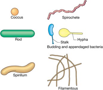
Cell Morphology
Prokaryotic cells exhibit a variety of shapes, which are important for identification and classification.
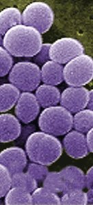
Cocci: Spherical; can occur singly (coccus), in pairs (diplococci), chains (streptococci), or clusters (staphylococci).
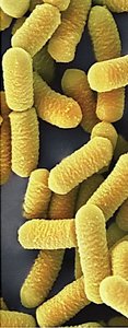
Bacilli: Rod-shaped; usually solitary (bacillus) or in chains (streptobacilli).
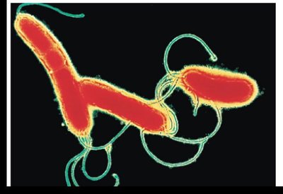
Spirilla/Spirillum: Spiral-shaped; range from comma-like to loose coils.

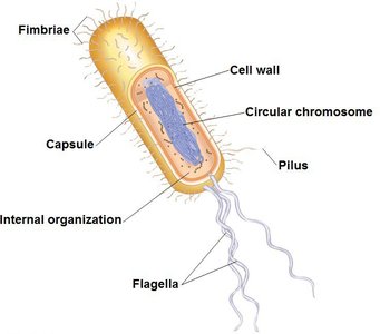
Typical Prokaryotic Cell Structure
Prokaryotic cells have a simple internal organization but possess specialized structures for survival and adaptation.
Cell wall: Provides shape, protection, and prevents lysis in hypotonic environments.
Capsule: Outer layer for protection and adherence.
Fimbriae and pili: Surface appendages for attachment and genetic exchange.
Flagella: Used for motility.
Circular chromosome: Contains genetic material.
Prokaryotic Cell Walls and Gram Staining
Composition and Function of Cell Walls
The cell wall is a critical feature of prokaryotic cells, maintaining shape, protecting the cell, and preventing osmotic lysis.
Bacterial cell walls: Contain peptidoglycan (murein), a network of sugar polymers (N-acetylglucosamine and N-acetylmuramic acid) cross-linked by polypeptides.
Archaeal cell walls: Lack peptidoglycan; contain glycoproteins, polysaccharides, or pseudomurein.
Eukaryotic cell walls: Made of cellulose (plants) or chitin (fungi).
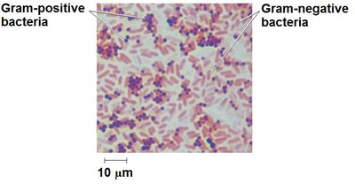
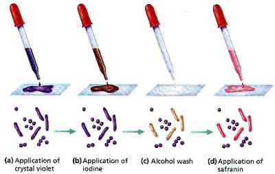
Gram Staining Procedure and Classification
The Gram stain is a differential staining technique that classifies bacteria based on cell wall composition:
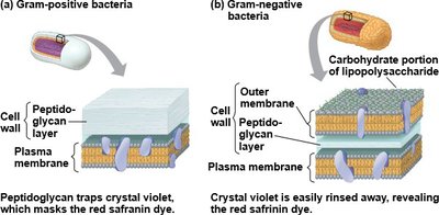
Gram-positive bacteria: Thick peptidoglycan layer, stain purple.
Gram-negative bacteria: Thin peptidoglycan layer, outer membrane with lipopolysaccharides (LPS), stain pink/red.
Biological and Medical Relevance
The lipid portion of LPS in Gram-negative bacteria is toxic and can trigger strong immune responses.
The outer membrane of Gram-negative bacteria provides resistance to certain antibiotics and host defenses.
Many antibiotics (e.g., penicillin) target peptidoglycan synthesis, making Gram-positive bacteria more susceptible.
Some Gram-positive bacteria have evolved resistance to multiple antibiotics, posing clinical challenges.
Additional Information
Endospores: Some bacteria can form resistant endospores to survive harsh conditions.
Modern uses of microbes: Biotechnology, genetic engineering, gene therapy, and environmental applications.
