 Back
BackProkaryotes: Structure, Function, and Diversity
Study Guide - Smart Notes
 Tailored notes based on your materials, expanded with key definitions, examples, and context.
Prokaryotes: Overview and Classification
Introduction to Prokaryotes
Prokaryotes are unicellular organisms that lack a membrane-bound nucleus and organelles. They are classified into two domains: Bacteria and Archaea. Prokaryotes are fundamental to the study of microbiology and play essential roles in ecosystems, health, and biotechnology. 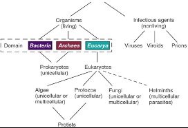
Cell Theory and Prokaryotic Cells
The cell theory, based on the work of Schleiden, Schwann, and Virchow, states:
All organisms are composed of cells.
Cells are the basic units of organization of all organisms.
Cells arise only from pre-existing cells.
Shared Features of Prokaryotic and Eukaryotic Cells
Both prokaryotic and eukaryotic cells possess:
Plasma membrane
Ribosomes
DNA organized in chromosomes
Prokaryotic Cell Morphologies and Arrangements
Common Cell Shapes
Prokaryotic cells exhibit several distinct morphologies, which are important for identification and classification. 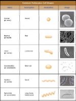
Coccus: Spherical
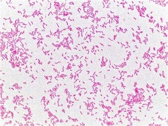
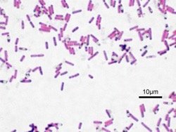
Bacillus: Rod-shaped
Vibrio: Curved rod
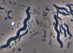
Spirillum: Spiral-shaped
Spirochete: Long, flexible spiral
Cell Arrangements
Prokaryotic cells can be found in various arrangements depending on their division patterns. 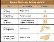
Coccus: Single spherical cell
Diplococcus: Pair of cocci
Tetrad: Group of four cocci
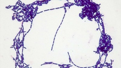
Streptococcus: Chain of cocci
Staphylococcus: Cluster of cocci
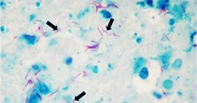
Microscopic Examples
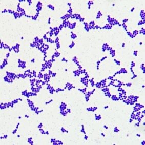
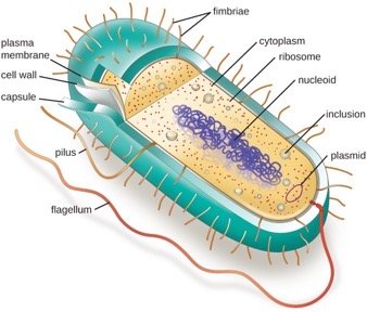
Bacterial Cell Structure
Essential Components
All bacterial cells contain:
Cell membrane
Cytoplasm
Ribosomes
Chromosome (DNA) in the nucleoid

Additional Structures
Most bacterial cells also possess:
Cell wall
Plasmids
Glycocalyx (slime layer or capsule)

Specialized Structures
Some bacteria have:
Second cell membrane
Flagella, pili, and/or fimbriae
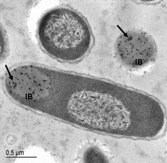
Inclusion bodies
Bacterial Cytoplasmic Structures
Nucleoid
Region containing the prokaryotic chromosome (double-stranded, supercoiled DNA)
Encodes for approximately 4,000 genes
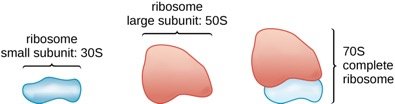
Ribosomes
Sites of protein synthesis
Composed of rRNA and proteins
Consist of a large (50S) and small (30S) subunit, forming a 70S ribosome
Protein Synthesis
Ribosomes catalyze the formation of peptide bonds between amino acids, resulting in polypeptide chains. 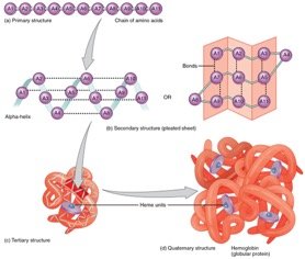
Plasmids
Small, circular DNA molecules
Encode non-essential functions (e.g., antibiotic resistance)
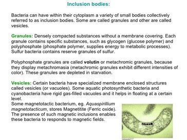
Inclusion Bodies
Serve as storage sites for nutrients and other substances.
Granules: Store phosphate, glycogen, or sulfur
Vesicles: Membrane-bound, may store gases or iron oxide
Cell Envelope: Structure and Function
Membrane Structure
The cell envelope consists of one or two plasma membranes and a cell wall.
Membranes are composed of phospholipids and proteins
Control transport in and out of the cell
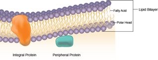
Fluid Mosaic Model
The membrane is a dynamic, non-rigid structure with amphipathic phospholipids forming a bilayer.
Integral proteins: Span the membrane, involved in transport and signaling
Peripheral proteins: Attached to the surface, with various functions

Membrane Transport
The plasma membrane is selectively permeable due to the amphipathic nature of phospholipids.
Controls movement of molecules
Maintains cellular homeostasis
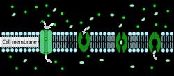
Transport Mechanisms
Diffusion
Simple diffusion allows small, non-charged molecules (e.g., O2, CO2) to move across the membrane down their concentration gradient.
Passive process; no energy required


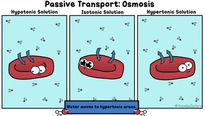
Osmosis
Osmosis is the diffusion of water across a selectively permeable membrane toward higher solute concentration.
Can cause osmotic pressure, affecting cell volume and function

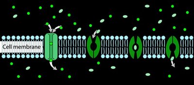
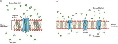
Facilitated Diffusion
Facilitated diffusion moves ions and molecules through membrane channels or carriers.
Passive; no energy required
Equilibrium is reached when solute concentration is equal on both sides
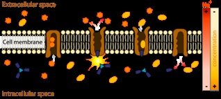
Active Transport
Active transport moves solutes against their concentration gradient (from low to high concentration).
Requires energy (usually ATP)
Involves specific transport proteins
Bacterial Appendages and Surface Structures
Glycocalyx
The glycocalyx is a coating of repeating polysaccharides or glycoproteins outside the cell membrane.
Functions: attachment, protection, prevention of water loss, nutrient trapping
Types: Slime layer (loose), Capsule (firm), S-layer (proteinaceous)
Cell Wall
The cell wall provides shape and structural support, preventing osmotic lysis.
Contains peptidoglycan, a polymer of NAG and NAM
Site of action for several antibiotics
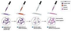
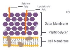
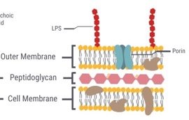
Gram Staining and Cell Wall Types
Gram Positive vs. Gram Negative
Gram staining differentiates bacteria based on cell wall structure.
Gram Positive: Thick peptidoglycan layer, teichoic acids, stains purple
Gram Negative: Thin peptidoglycan layer, outer membrane, stains pink
Atypical Cell Walls
Mycolic acid: Found in Mycobacterium and Nocardia; acid-fast staining required
Contributes to resistance and pathogenicity
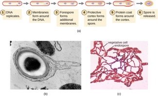
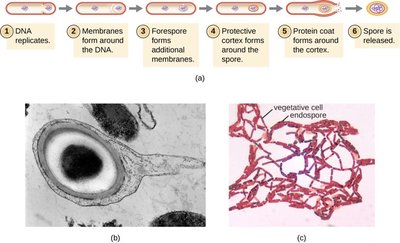
Bacterial Endospores
Endospore Formation and Germination
Some bacteria form endospores under nutrient-limited conditions.
Vegetative state: Metabolically active
Endospore: Dormant, highly resistant to heat, chemicals, radiation, and desiccation
Sporulation: Formation of endospore
Germination: Return to vegetative state when conditions improve
Bacterial Appendages
Flagella
Flagella are responsible for motility in aqueous environments.
Arrangements: Monotrichous (single), Amphitrichous (both ends), Lophotrichous (tuft at one end), Peritrichous (all over)
Movement: "Tumble and run" mechanism; chemotaxis and phototaxis
Pili and Fimbriae
Composed of pilin protein
Functions: Attachment, communication, conjugation (genetic exchange)
Archaea: Unique Features
Extremophiles
Archaea are often found in extreme environments.
Thermophiles: High temperatures
Halophiles: High salt or acid concentrations
Methanogens: Produce methane, anaerobic
Archaeal Cell Walls
May be composed of polysaccharides or pure protein
Lack true peptidoglycan
Some lack a cell wall entirely
Summary Table: Gram Positive vs. Gram Negative Cell Walls
Feature | Gram Positive | Gram Negative |
|---|---|---|
Peptidoglycan Thickness | Thick (20–80 nm) | Thin (1–3 nm) |
Teichoic Acids | Present | Absent |
Outer Membrane | Absent | Present |
Stain Color | Purple | Pink |
Additional info: This guide expands on the original notes with definitions, examples, and academic context to support foundational understanding of prokaryotic cell biology.
