 Back
BackProkaryotic Cells: Structure, Function, and Laboratory Techniques
Study Guide - Smart Notes
 Tailored notes based on your materials, expanded with key definitions, examples, and context.
Prokaryotic and Eukaryotic Cells: An Overview
Major Types of Cells
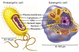
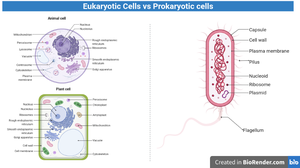
Cells are the fundamental units of life and can be classified into two major types: prokaryotic cells and eukaryotic cells. Understanding their differences is essential for studying cellular biology.
Prokaryotic Cells: Generally unicellular, small (0.1–10 μm), and structurally simpler. Examples include bacteria and archaea.
Eukaryotic Cells: Can be unicellular or multicellular, larger (10–100 μm), and more complex. Examples include plants, animals, fungi, and protists.
Genetic Material Organization
The organization of genetic material is a key distinction between prokaryotes and eukaryotes.
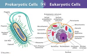
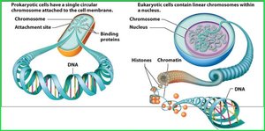
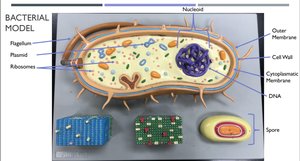
Prokaryotes: Possess a single, circular DNA molecule located in a region called the nucleoid. DNA is not enclosed by a membrane.
Eukaryotes: Contain multiple, linear chromosomes housed within a membrane-bound nucleus.
Cellular Organelles
Organelles are specialized structures within cells that perform distinct functions.
Prokaryotes: Lack membrane-bound organelles but contain ribosomes for protein synthesis.
Eukaryotes: Possess numerous membrane-bound organelles (e.g., mitochondria, endoplasmic reticulum, Golgi apparatus) and ribosomes.
Storage of Genetic Material
Prokaryotes: DNA is found in the cytoplasm within the nucleoid region.
Eukaryotes: DNA is stored as linear chromosomes inside a membrane-bound nucleus, separated from the cytoplasm.
Nutrition in Prokaryotes
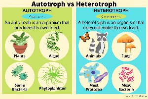
Autotrophs vs. Heterotrophs
Prokaryotes can obtain energy and carbon in different ways, classified as autotrophs or heterotrophs.
Autotrophs: Organisms that produce their own food, typically using sunlight (photosynthesis) or inorganic chemicals (chemosynthesis).
Heterotrophs: Organisms that obtain food by consuming other organisms or organic matter.
Bacterial Morphology and Identification
Common Shapes of Bacteria
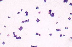
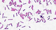
Bacteria are classified by their morphology (shape):
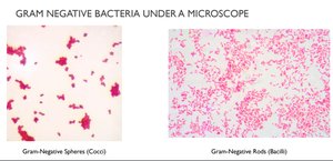
Coccus (cocci): Spherical-shaped bacteria.
Bacillus (bacilli): Rod-shaped bacteria.
Spirillum (spirilli): Spiral or helical-shaped bacteria.
Bacterial Cell Walls and Gram Staining
Structure of Bacterial Cell Walls
Bacterial cell walls are essential for maintaining cell shape and integrity. They are classified as Gram-positive or Gram-negative based on their structure and staining properties.
Feature | Gram-Positive | Gram-Negative |
|---|---|---|
Peptidoglycan Layer | Thick | Thin |
Lipopolysaccharide Layer | Absent | Present |
Outer Membrane | Absent | Present |
Stain Color | Purple | Pink |
Peptidoglycan is a polymer of sugars and amino acids that forms a mesh-like layer outside the plasma membrane.
Gram Staining Procedure
Gram staining differentiates bacteria based on cell wall structure using a series of dyes and reagents:
Crystal Violet: Primary stain; stains all cells purple.
Gram's Iodine: Mordant; forms a complex with crystal violet, fixing the dye in Gram-positive cells.
Ethanol: Decolorizing agent; dehydrates thick peptidoglycan in Gram-positive cells (retains dye), dissolves outer membrane in Gram-negative cells (dye washes out).
Safranin: Counterstain; stains Gram-negative cells pink, Gram-positive cells remain purple.
Gram-positive bacteria appear purple, while Gram-negative bacteria appear pink under the microscope.
Cyanobacteria
Characteristics of Cyanobacteria
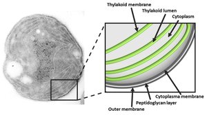
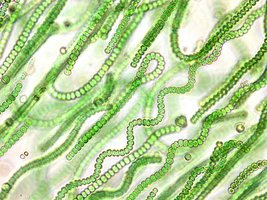
Cyanobacteria are photosynthetic prokaryotes, often called "blue-green algae" due to their pigmentation. They are important for oxygen production and nitrogen fixation in aquatic environments.
Contain chlorophyll a (unlike most other photosynthetic prokaryotes, which have bacteriochlorophyll).
Lack chloroplasts; photosynthesis occurs in thylakoid membranes within the cytoplasm.
Often form linear filaments and do not fit standard bacterial morphology terms.
Phycobilins give them a blue-green hue.
Laboratory Techniques: Culturing and Identifying Bacteria
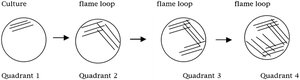
Isolation Streak Plate (4-Quadrant Streak Plate)
This technique is used to isolate individual bacterial colonies from a mixed sample by streaking bacteria across four quadrants of an agar plate, decreasing the concentration with each quadrant.
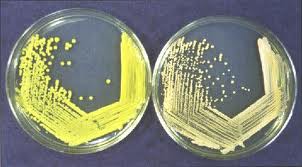
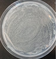
Spread Plate (Lawn Plate)
The spread plate technique creates a uniform layer of bacterial growth across the surface of the agar, useful for quantifying bacteria or testing antibiotic sensitivity.
Laboratory Safety and Best Practices
Always label agar plates with your name, lab section, date, and type of plate. Store agar side up and wrap in parafilm.
Slides must be air-dried before heat fixing; gently pass glass through the flame to avoid breakage.
Dispose of used slides and plates in designated discard bins.
Additional info: Proper aseptic technique is essential to prevent contamination and ensure accurate results in microbiology labs.
