 Back
BackBio 100 Lec Chapter 5 Part 3
Study Guide - Smart Notes
 Tailored notes based on your materials, expanded with key definitions, examples, and context.
Bio 100 Lec Chapter 5 (Part 3)
The Structure and Function of Large Biological Molecules
Protein Structure: Levels and Stabilizing Interactions
Proteins are complex macromolecules whose function is determined by their structure, which is organized into four hierarchical levels: primary, secondary, tertiary, and quaternary. Each level is stabilized by specific types of chemical interactions.
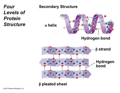
Secondary Structure
The secondary structure of proteins arises from hydrogen bonding between the repeating constituents of the polypeptide backbone. The two main types are:
Alpha helix (α helix): A spiral structure stabilized by intramolecular hydrogen bonds between the amino and carboxyl groups of the same polypeptide chain.
Beta pleated sheet (β sheet): A folded structure formed by hydrogen bonds between segments of the polypeptide backbone, which can be parallel or antiparallel. Beta sheets can participate in both intramolecular and intermolecular bonding.
Hydrogen bonds: These bonds occur at regular intervals, creating patterns that stabilize the secondary structure.
Example: The formation of α helices and β sheets depends on the amino acid sequence, though the specific prediction of which amino acids form these structures is beyond introductory biology.
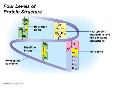
Tertiary Structure
The tertiary structure is the overall three-dimensional shape of a polypeptide, resulting from interactions among the side chains (R groups) of the amino acids. These interactions include:
Hydrogen bonds: Occur between polar side chains, allowing distant residues to interact after folding.
Ionic bonds: Form between charged side chains (acidic and basic), and are sensitive to environmental pH.
Hydrophobic interactions: Nonpolar side chains cluster away from water, driving protein folding.
Van der Waals interactions: Weak, transient attractions between nonpolar molecules due to temporary dipole moments.
Disulfide bridges: Covalent bonds between sulfhydryl groups of cysteine residues, providing strong stabilization.
Example: Insulin is stabilized by intermolecular disulfide bridges between its two polypeptide chains.
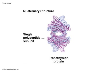
Quaternary Structure
Quaternary structure arises when two or more polypeptide chains (subunits) assemble into a functional protein. The same forces stabilizing tertiary structure also stabilize quaternary structure. Multimeric proteins may have identical or different subunits.
Example: Transthyretin protein consists of four polypeptide subunits, arranged in pairs.
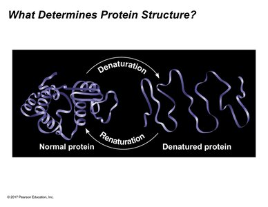
Protein Stability and Denaturation
Protein function depends on its correct folding and structure. Environmental factors such as pH, salinity, and temperature can disrupt stabilizing interactions, leading to denaturation—the loss of native structure and biological activity. Denatured proteins retain their primary sequence but lose higher-order structure. Under optimal conditions, some proteins can renature, regaining function.
Denaturation: Loss of secondary, tertiary, and quaternary structure due to environmental changes.
Renaturation: Restoration of native structure and function under favorable conditions.
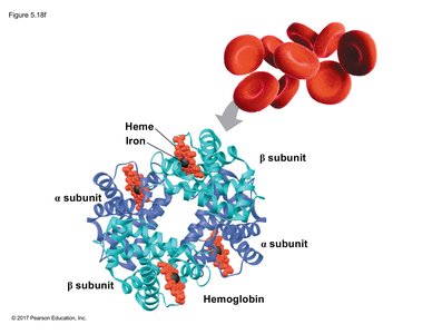
Form and Function: Hemoglobin and Sickle Cell Disease
Hemoglobin is a tetrameric protein found in red blood cells, composed of two alpha and two beta subunits. Its function is to transport oxygen efficiently. The primary structure of hemoglobin is critical for its proper folding and function.
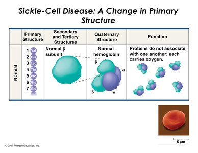
Normal hemoglobin: Subunits do not aggregate; red blood cells are biconcave and efficient in oxygen transport.
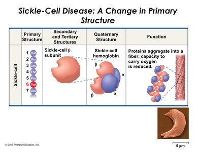
Sickle Cell Disease
Sickle cell disease results from a single amino acid substitution in the beta subunit of hemoglobin (glutamic acid replaced by valine). This change alters the protein's folding, causing aggregation and fiber formation, which distorts red blood cell shape and impairs oxygen transport.
Primary Structure | Secondary and Tertiary Structures | Quaternary Structure | Function |
|---|---|---|---|
Normal: Glu at position 6 | Normal β subunit | Normal hemoglobin (α2β2) | Proteins do not associate; efficient oxygen transport |
Sickle-cell: Val at position 6 | Sickle-cell β subunit | Sickle-cell hemoglobin (α2β2) | Proteins aggregate; reduced oxygen transport |
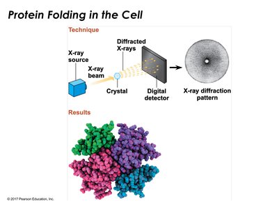
Protein Folding Techniques
Determining protein structure is complex. X-ray crystallography is a key technique, where a crystallized protein is irradiated with X-rays, producing a diffraction pattern that reveals atomic positions and enables construction of a three-dimensional model.
Crystallization: Produces defect-free crystals for accurate data.
X-ray diffraction: Analyzes spot patterns to infer atomic arrangement.
Nucleic Acids: Structure and Function
Overview and Biological Role
Nucleic acids are macromolecules responsible for storing, transmitting, and expressing hereditary information. Genes, composed of DNA, encode the amino acid sequence of polypeptides. There are two major types of nucleic acids: DNA and RNA.
Gene: A unit of inheritance made of DNA.
Nucleic acid: Polymer of nucleotide monomers.
Central Dogma of Molecular Biology
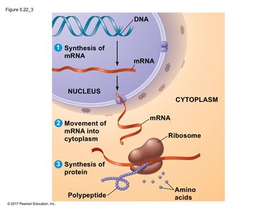
The flow of genetic information follows the central dogma: DNA is transcribed into RNA, which is then translated into protein. Messenger RNA (mRNA) carries genetic instructions from the nucleus to the cytoplasm, where ribosomes synthesize polypeptides.
Transcription: DNA → RNA (mRNA)
Translation: RNA → Protein
Nucleotide Structure and Polynucleotide Formation
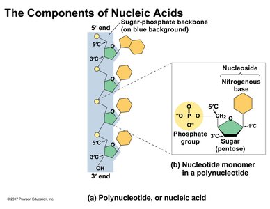
Nucleic acids are polymers called polynucleotides, composed of nucleotide monomers. Each nucleotide consists of a nitrogenous base, a pentose sugar (ribose or deoxyribose), and a phosphate group. Nucleotides are linked by phosphodiester bonds, forming a sugar-phosphate backbone.
Nucleotide: Nitrogenous base + sugar + phosphate group
Nucleoside: Nitrogenous base + sugar (no phosphate)
Phosphodiester linkage: Connects 5' carbon of one sugar to 3' carbon of the next
Nitrogenous Bases
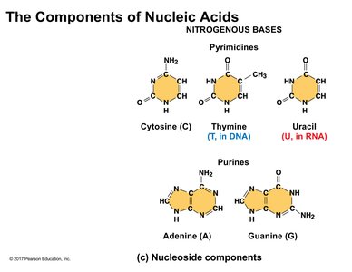
Nitrogenous bases are classified into two families:
Pyrimidines: Single six-membered ring (cytosine, thymine, uracil)
Purines: Two fused rings (adenine, guanine)
Thymine: Found in DNA only
Uracil: Found in RNA only
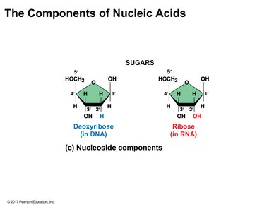
Pentose Sugars
The pentose sugar distinguishes DNA from RNA:
Deoxyribose: Found in DNA; has a hydrogen at the 2' position
Ribose: Found in RNA; has a hydroxyl group at the 2' position
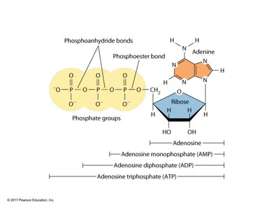
Adenosine Triphosphate (ATP): A Nucleotide Example
ATP is a nucleotide composed of adenine (nitrogenous base), ribose (sugar), and three phosphate groups. The energy stored in ATP is primarily in the phosphoanhydride bonds between phosphate groups.
Phosphoanhydride bond: High-energy bond between phosphate groups
Phosphoester bond: Bond between phosphate and sugar
Polynucleotide Directionality
Polynucleotides have intrinsic directionality, synthesized from the 5' end to the 3' end. The backbone consists of alternating sugar and phosphate groups, while the sequence of nitrogenous bases encodes genetic information.
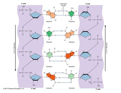
Double-Stranded DNA and Base Pairing
DNA typically exists as a double-stranded molecule, stabilized by complementary base pairing between nitrogenous bases:
Adenine (A) pairs with Thymine (T): Two hydrogen bonds
Cytosine (C) pairs with Guanine (G): Three hydrogen bonds
Antiparallel orientation: Strands run in opposite directions (5' to 3' and 3' to 5')
Backbone: Polar, faces outward; bases are hydrophobic, face inward
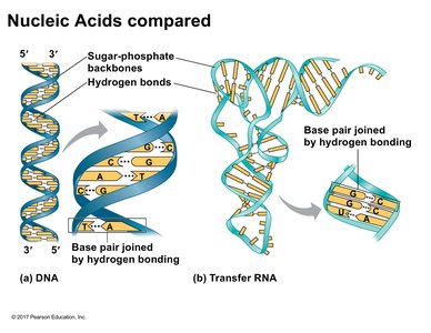
Comparison of DNA and RNA
DNA is usually double-stranded, while RNA is typically single-stranded but can form regions of complementary base pairing. Transfer RNA (tRNA) folds upon itself, creating antiparallel regions stabilized by hydrogen bonds. Some viruses possess double-stranded RNA.
DNA: Double-stranded, uses thymine
RNA: Single-stranded, uses uracil
tRNA: Example of RNA folding and base pairing
Additional info: These notes expand on the original lecture and slide content, providing definitions, examples, and context for protein and nucleic acid structure, as well as their biological significance.
