 Back
BackProtein Structure and Enzyme Function: Study Notes for Principles of Biology
Study Guide - Smart Notes
 Tailored notes based on your materials, expanded with key definitions, examples, and context.
Protein Structure and Function
Overview of Protein Function
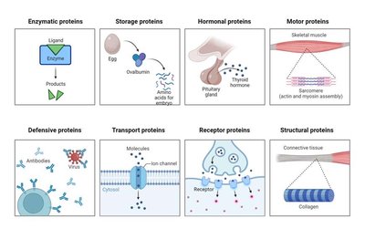
Proteins are essential biomolecules that perform a wide variety of functions in living organisms. Their structure determines their function, and they are involved in catalysis, transport, defense, structure, signaling, and movement.
Enzymatic proteins: Catalyze biochemical reactions (e.g., catalase).
Storage proteins: Store amino acids or other substances (e.g., ovalbumin in eggs).
Hormonal proteins: Coordinate organismal activities (e.g., insulin, thyroid hormone).
Motor proteins: Enable movement (e.g., actin and myosin in muscles).
Defensive proteins: Protect against disease (e.g., antibodies).
Transport proteins: Move substances across membranes (e.g., ion channels).
Receptor proteins: Receive and transmit signals (e.g., neurotransmitter receptors).
Structural proteins: Provide support (e.g., collagen in connective tissue).
Amino Acid Structure
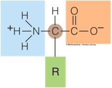
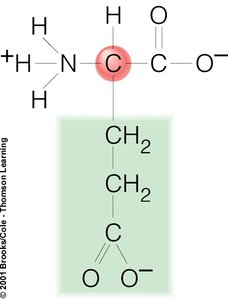
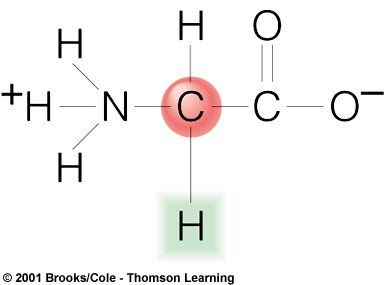
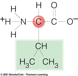
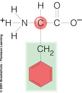
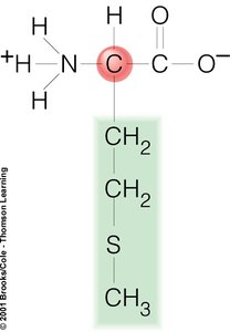
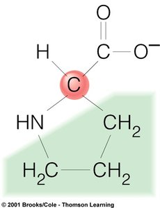
Amino acids are the building blocks of proteins. Each amino acid contains a central carbon atom (the alpha carbon) bonded to an amino group, a carboxyl group, a hydrogen atom, and a variable side chain (R group). There are 20 different amino acids, each with a unique R group that determines its properties.
General structure: Central carbon, amino group, carboxyl group, hydrogen, and R group.
R group: Determines the identity and chemical behavior of the amino acid.
Examples of Amino Acids
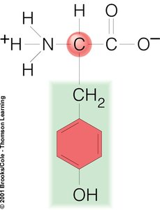
Tyrosine (Tyr): Contains a phenol group.
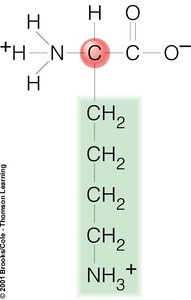
Lysine (Lys): Contains a long aliphatic chain ending in an amino group.
Glutamate (Glu): Contains a carboxyl group in its side chain.
Glycine (Gly): Has a single hydrogen as its R group.
Valine (Val): Has a branched aliphatic side chain.
Phenylalanine (Phe): Contains a benzyl side chain.
Methionine (Met): Contains a sulfur atom in its side chain.
Proline (Pro): Has a unique cyclic structure.
Levels of Protein Structure
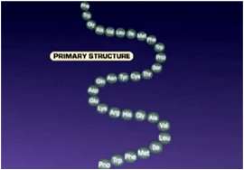
Primary Structure
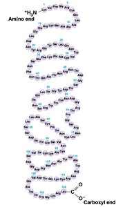
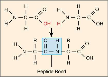
The primary structure of a protein is its unique sequence of amino acids, linked together by covalent peptide bonds. The sequence is determined by the genetic code and is written from the amino (N) terminus to the carboxyl (C) terminus.
Peptide bond: Covalent bond formed between the carboxyl group of one amino acid and the amino group of the next.
Directionality: N-terminus to C-terminus.
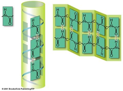
Secondary Structure
The secondary structure refers to local folding patterns within a polypeptide, stabilized by hydrogen bonds. The most common types are the alpha helix and beta pleated sheet.
Alpha helix: Right-handed coil stabilized by hydrogen bonds between every fourth amino acid.
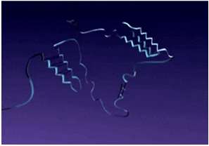
Beta pleated sheet: Sheet-like structure formed by hydrogen bonds between parallel or antiparallel strands.


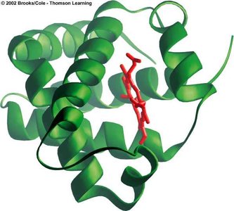
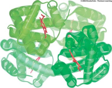
Tertiary Structure
The tertiary structure is the overall three-dimensional shape of a single polypeptide chain, stabilized by interactions between R groups, including hydrogen bonds, ionic bonds, hydrophobic interactions, and disulfide bridges. This level of structure determines the protein's functional conformation.
Globular shape: Many proteins fold into compact, globular forms.
Stabilizing interactions: Covalent, ionic, hydrogen bonds, and hydrophobic interactions between R groups.


Quaternary Structure
Some proteins are composed of more than one polypeptide chain. The quaternary structure refers to the arrangement and interaction of these multiple subunits in a functional protein complex.
Subunit assembly: Multiple polypeptide chains (subunits) associate to form a functional protein.
Examples: Hemoglobin (four subunits), catalase (multiple subunits).


Enzymes and Chemical Reactions
Enzyme Structure and Function
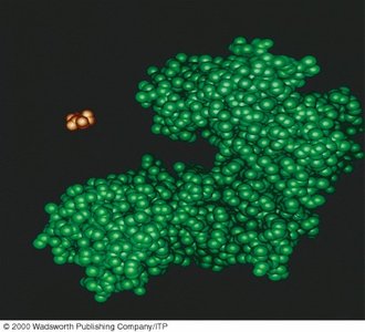
Enzymes are biological catalysts, usually proteins, that speed up chemical reactions by lowering the activation energy required. Each enzyme is specific to its substrate due to the unique shape of its active site.
Active site: Region on the enzyme where the substrate binds and the reaction occurs.
Specificity: Enzymes are highly specific for their substrates.
Chemical Reactions: Synthesis and Breakdown
Chemical reactions in cells can be classified as synthesis (anabolic) or breakdown (catabolic) reactions.
Synthesis (anabolic): Two or more reactants combine to form a larger product. Example:
Breakdown (catabolic): A reactant is broken down into two or more products. Example:

Catalase and Enzyme Specificity
Catalase is an enzyme that catalyzes the breakdown of hydrogen peroxide into water and oxygen. Enzymes like catalase are highly specific, binding only to their particular substrate.
Reaction:
Substrate: Hydrogen peroxide ()
Products: Water () and oxygen ()
Specificity: Catalase only acts on hydrogen peroxide, not other molecules.
Experimental Design and Analysis
Investigating Enzyme Activity
Experiments with catalase often involve measuring the rate of hydrogen peroxide breakdown by observing the production of oxygen (e.g., foam height in a test tube). Key experimental considerations include controls, variables, and sample size.
Experimental variable: The factor being tested (e.g., presence of catalase).
Control: A sample without the experimental variable (e.g., tube without catalase).
Sample size: Number of replicates for reliability.
Key Experimental Questions
Does catalase increase the rate of hydrogen peroxide breakdown?
Has all the substrate been converted in the reaction?
Is the enzyme still functional after the reaction?
These questions are addressed by comparing experimental and control tubes, observing foam production, and testing for remaining substrate or enzyme activity.
Laboratory Techniques and Clean-Up
Best Practices
Label tubes clearly.
Measure foam height accurately using millimeter rulers or pipettes.
Mix tubes thoroughly before measuring.
Clean all equipment and workspaces after the experiment.
Summary Table: Levels of Protein Structure
Level | Description | Bonds/Interactions | Example |
|---|---|---|---|
Primary | Sequence of amino acids | Peptide bonds | Insulin chain |
Secondary | Local folding (alpha helix, beta sheet) | Hydrogen bonds | Alpha helix in keratin |
Tertiary | 3D shape of polypeptide | Hydrogen, ionic, disulfide, hydrophobic | Myoglobin |
Quaternary | Assembly of multiple polypeptides | Same as tertiary (between subunits) | Hemoglobin |
Additional info: The notes above expand on the brief points in the original material, providing definitions, examples, and context for each concept. The images included are only those directly relevant to the explanation of protein structure and enzyme function.
