 Back
BackProtein Structure and Enzyme Function: Study Notes for Principles of Biology
Study Guide - Smart Notes
 Tailored notes based on your materials, expanded with key definitions, examples, and context.
Protein Structure and Function
Overview of Protein Function
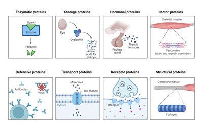
Proteins are essential biomolecules that perform a wide variety of functions in living organisms. Their structure determines their function, and they are involved in catalysis, transport, signaling, structure, and defense.
Enzymatic proteins: Catalyze biochemical reactions (e.g., catalase).
Storage proteins: Store amino acids or other substances (e.g., ovalbumin in eggs).
Hormonal proteins: Coordinate organismal activities (e.g., insulin, thyroid hormone).
Motor proteins: Enable movement (e.g., actin and myosin in muscles).
Defensive proteins: Protect against disease (e.g., antibodies).
Transport proteins: Move substances across membranes (e.g., ion channels).
Receptor proteins: Receive and transmit signals (e.g., neurotransmitter receptors).
Structural proteins: Provide support (e.g., collagen in connective tissue).
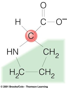
Amino Acid Structure
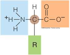
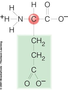
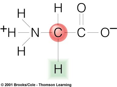
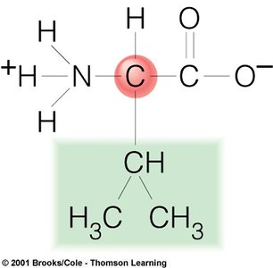
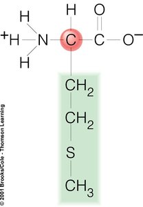
Proteins are polymers of amino acids. Each amino acid has a central carbon (alpha carbon) bonded to an amino group, a carboxyl group, a hydrogen atom, and a variable R group (side chain). The R group determines the properties and identity of each amino acid.
20 standard amino acids are used to build proteins.
The R group (side chain) is unique for each amino acid and determines its chemical behavior.
Examples of Amino Acids
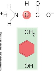
Tyrosine (Tyr): Contains a phenol group.
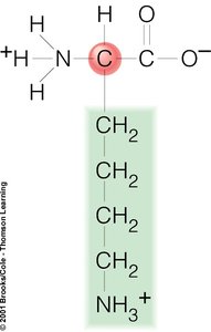
Lysine (Lys): Contains a long aliphatic chain ending with an amino group.
Glutamate (Glu): Contains a carboxyl group in its side chain.
Glycine (Gly): Has a single hydrogen as its side chain.
Valine (Val): Has a branched aliphatic side chain.
Phenylalanine (Phe): Contains a benzyl side chain.
Methionine (Met): Contains a sulfur atom in its side chain.
Proline (Pro): Has a unique cyclic structure.

Levels of Protein Structure
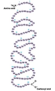
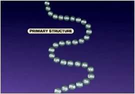
Primary Structure
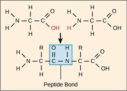
The primary structure of a protein is the unique sequence of amino acids in a polypeptide chain. This sequence is determined by the gene encoding the protein and is held together by covalent peptide bonds.
Peptide bonds form between the carboxyl group of one amino acid and the amino group of the next.
The sequence is read from the N-terminus (amino end) to the C-terminus (carboxyl end).
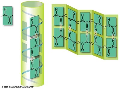
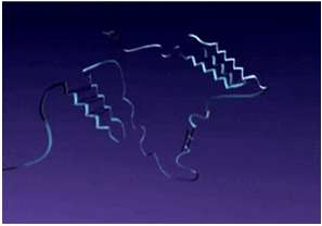
Secondary Structure
The secondary structure refers to local folding patterns within a polypeptide, stabilized by hydrogen bonds. The most common types are the alpha helix and beta pleated sheet.
Alpha helix: A right-handed coil stabilized by hydrogen bonds between every fourth amino acid.
Beta pleated sheet: Sheet-like arrangement formed by hydrogen bonds between segments of the polypeptide chain.
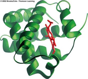
Tertiary Structure
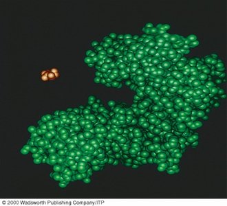
The tertiary structure is the overall three-dimensional shape of a single polypeptide chain, resulting from interactions between R groups. These include hydrogen bonds, ionic bonds, hydrophobic interactions, and disulfide bridges.
Globular shape is common for many functional proteins.
Stabilized by interactions among side chains (R groups).

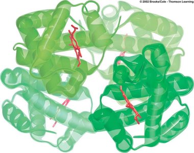
Quaternary Structure
Some proteins are composed of more than one polypeptide chain. The quaternary structure describes the arrangement and interaction of these subunits.
Examples: Hemoglobin (four subunits), catalase (multiple subunits).
Stabilized by the same types of interactions as tertiary structure.

Enzymes and Chemical Reactions
Enzyme Structure and Function
Enzymes are biological catalysts, usually proteins, that speed up chemical reactions without being consumed. They are highly specific for their substrates and lower the activation energy required for reactions.
Active site: The region of the enzyme where the substrate binds and the reaction occurs.
Specificity: Each enzyme only catalyzes a specific reaction or set of reactions.
Chemical Reactions: Anabolic and Catabolic
Chemical reactions in cells can be classified as anabolic (building molecules) or catabolic (breaking down molecules).
Synthesis (anabolic) reaction: Two or more reactants combine to form a product. Example:
Breakdown (catabolic) reaction: A reactant is broken down into two or more products. Example:

Catalase and Hydrogen Peroxide Breakdown
Catalase is an enzyme that catalyzes the breakdown of hydrogen peroxide (H2O2) into water and oxygen, preventing cellular damage from this reactive molecule.
Reaction:
Importance: Protects cells from oxidative damage.
Enzyme Specificity
Enzymes are highly specific, meaning each enzyme only binds to a particular substrate and catalyzes a specific reaction. This specificity is due to the precise shape and chemical environment of the enzyme's active site.
Substrate: The molecule upon which an enzyme acts.
Product: The molecule(s) produced by the enzyme-catalyzed reaction.
Experimental Design: Enzyme Activity
Investigating Catalase Activity
Experiments can be designed to test the effect of catalase on hydrogen peroxide breakdown, the completeness of substrate conversion, and the functionality of the enzyme after the reaction.
Measure the rate of reaction by observing the height of foam produced (oxygen release).
Use controls to determine the experimental variable and sample size.
Test for remaining substrate or enzyme by adding more reactants or enzyme to the reaction mixture.
Sample Experimental Questions
Does catalase increase the rate of hydrogen peroxide breakdown?
Has all the substrate been converted in the reaction?
Is the enzyme still functional after the reaction?
Laboratory Techniques and Clean-Up
Best Practices
Proper laboratory technique is essential for accurate results and safety. This includes labeling tubes, measuring accurately, mixing samples, and cleaning up thoroughly after experiments.
Label all tubes clearly.
Measure foam height in millimeters using pipettes.
Mix tubes at a 45-degree angle and flick gently.
Clean all equipment and wash hands after the experiment.
