 Back
BackProtein Structure and Function: Study Notes
Study Guide - Smart Notes
 Tailored notes based on your materials, expanded with key definitions, examples, and context.
Protein Structure and Function
3.1 Amino Acids and Their Polymerization
Amino acids are the building blocks of proteins, each sharing a common core structure but differing in their side chains (R-groups). The chemical properties of amino acids are determined by their side chains, which influence protein structure and function.
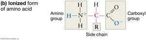
Core Structure: Each amino acid consists of a central (alpha) carbon atom bonded to a hydrogen atom, an amino group (–NH2), a carboxyl group (–COOH), and a variable R-group (side chain).
Ionization in Water: In aqueous environments, the amino group tends to gain a proton (becoming –NH3+), and the carboxyl group loses a proton (becoming –COO–), which helps amino acids stay in solution and affects their reactivity.
R-Groups: The R-group is the variable part of the amino acid and determines its unique properties. Some R-groups contain functional groups that can participate in chemical reactions, while others are composed solely of carbon and hydrogen.
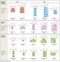
Classification by R-Group: Amino acids are classified based on the polarity and charge of their R-groups:
Charged (acidic or basic): Side chains with a negative charge are acidic (lost a proton), while those with a positive charge are basic (gained a proton).
Uncharged Polar: Side chains with an oxygen atom (highly electronegative) form polar covalent bonds.
Nonpolar: Side chains lacking charge or oxygen are nonpolar and hydrophobic.
Solubility: Polar and charged R-groups are hydrophilic and interact with water, while nonpolar R-groups are hydrophobic and do not form hydrogen bonds with water.
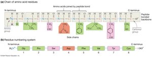
Peptide Bond Formation: Amino acids link via condensation reactions, forming covalent peptide bonds between the carboxyl group of one amino acid and the amino group of another. This process releases a molecule of water.

Peptide-Bonded Backbone: The resulting polypeptide has directionality (N-terminus to C-terminus), R-group orientation (side chains extend outward), and flexibility (rotation possible around single bonds adjacent to the peptide bond).
Oligopeptides and Polypeptides: Chains with fewer than 50 amino acids are called oligopeptides or peptides; those with more than 50 are polypeptides. A protein is a complete, functional polypeptide.
3.2 What Do Proteins Look Like?
Protein structure is hierarchical and determines function. The diversity of protein shapes and sizes arises from the sequence and properties of amino acids.
Primary Structure: The unique sequence of amino acids in a protein. Even a single amino acid change can drastically alter protein function (e.g., sickle cell hemoglobin).
Secondary Structure: Local folding patterns stabilized by hydrogen bonds between backbone atoms. The two main types are:
Alpha-helix (α-helix): A coiled structure stabilized by hydrogen bonds.
Beta-pleated sheet (β-sheet): Sheet-like structures formed by hydrogen bonding between different segments of the polypeptide chain.
Tertiary Structure: The overall three-dimensional shape of a polypeptide, resulting from interactions among R-groups, including:
Hydrogen bonding
Hydrophobic interactions
van der Waals interactions
Covalent bonding (e.g., disulfide bridges)
Ionic bonding
Quaternary Structure: The arrangement of multiple polypeptide subunits in a protein. Subunits may be identical (homodimers) or different (heterodimers). Some proteins form large complexes called macromolecular machines (e.g., ribosomes).
Level of Structure | Description | Stabilizing Bonds/Interactions |
|---|---|---|
Primary | Sequence of amino acids | Peptide bonds |
Secondary | Local folding (α-helix, β-sheet) | Hydrogen bonds |
Tertiary | Three-dimensional shape | R-group interactions (hydrogen, ionic, hydrophobic, van der Waals, covalent) |
Quaternary | Multiple polypeptide subunits | Interactions between subunits |
3.3 Folding and Function
Proper protein folding is essential for function. Folding is often spontaneous and results in a more stable, lower-energy state. Misfolded proteins can lose function or become harmful.
Molecular Chaperones: Specialized proteins that assist in the proper folding of other proteins and prevent inappropriate interactions. Example: Heat shock proteins (e.g., Hsp90).
Protein Flexibility: Many proteins are dynamic and can adopt multiple conformations until stabilized by binding to other molecules.
Prions: Misfolded proteins that can induce normal proteins to adopt the misfolded, disease-causing conformation (e.g., prion protein in mad cow disease).
3.4 Protein Functions Are as Diverse as Protein Structures
Proteins perform a wide variety of functions in cells, each dependent on their unique structure.
Catalysis: Enzymes are proteins that speed up chemical reactions by lowering activation energy. They bind substrates at specific active sites.
Structure: Proteins provide structural support to cells and tissues (e.g., collagen, keratin).
Movement: Motor proteins move cells or molecules within cells (e.g., myosin, kinesin).
Signaling: Proteins transmit signals between cells (e.g., hormones, receptors).
Transport: Proteins transport molecules across membranes or throughout the body (e.g., hemoglobin, channel proteins).
Defense: Antibodies are proteins that recognize and neutralize pathogens.
Enzyme Catalysis Example: Enzymes hold substrates in a precise orientation at the active site, facilitating the chemical reaction.
