 Back
Proteins and Nucleic Acids: Structure, Function, and Biological Importance
Study Guide - Smart Notes
 Tailored notes based on your materials, expanded with key definitions, examples, and context.
Proteins: Structure and Denaturation
Levels of Protein Structure
Proteins are complex biological macromolecules essential for structure, function, and regulation of the body's tissues and organs. Their structure is organized into four hierarchical levels:
Primary structure: The unique sequence of amino acids in a polypeptide chain, held together by peptide bonds.
Secondary structure: Local folding into structures such as alpha-helices and beta-pleated sheets, stabilized by hydrogen bonds.
Tertiary structure: The overall three-dimensional shape of a single polypeptide, stabilized by interactions including hydrogen bonds, ionic bonds, disulfide bridges, and hydrophobic interactions.
Quaternary structure: The association of multiple polypeptide chains into a functional protein complex.
Denaturation of Proteins
Denaturation refers to the loss of a protein's native structure due to the disruption of non-covalent interactions and disulfide bonds. It is generally easier to denature the secondary, tertiary, and quaternary structures than the primary structure, as the primary structure is maintained by strong covalent peptide bonds, while higher-order structures rely on weaker interactions.
Effect of pH: Changes in pH can disrupt ionic bonds and hydrogen bonds, leading to denaturation of secondary, tertiary, and quaternary structures.
Bonds affected: Hydrogen bonds, ionic bonds, and disulfide bridges are most susceptible to disruption by changes in pH or temperature.
Amino Acid Structure
Each amino acid contains a central (alpha) carbon atom bonded to four different groups:
An amino group (–NH2)
A carboxyl group (–COOH)
A hydrogen atom
A variable side chain (R group), which determines the properties and classification of the amino acid
The R groups can be nonpolar, polar, acidic, or basic, influencing the protein's structure and function.
Nucleic Acids: DNA and RNA
Introduction to Nucleic Acids
Nucleic acids are the molecules responsible for the storage, transmission, and expression of genetic information. The two main types are deoxyribonucleic acid (DNA) and ribonucleic acid (RNA). DNA is the hereditary material in most organisms, while RNA plays various roles in gene expression and protein synthesis.
Function: Nucleic acids code for the amino acid sequence in proteins, serving as the chemical link between generations.
Location: DNA is found in chromosomes and is passed from parent to offspring.
Structure of Nucleic Acids
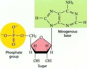
The basic building block (monomer) of nucleic acids is the nucleotide. Each nucleotide consists of three components:
A pentose sugar (deoxyribose in DNA, ribose in RNA)
A phosphate group
A nitrogenous base
DNA vs. RNA
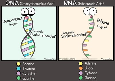
DNA and RNA differ in their sugar component, nitrogenous bases, and structure:
Feature | DNA | RNA |
|---|---|---|
Sugar | Deoxyribose | Ribose |
Nitrogenous Bases | Adenine, Thymine, Cytosine, Guanine | Adenine, Uracil, Cytosine, Guanine |
Strandedness | Double-stranded helix | Single-stranded |
Nitrogenous Bases
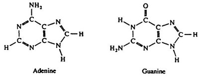
Nitrogenous bases are classified into two groups:
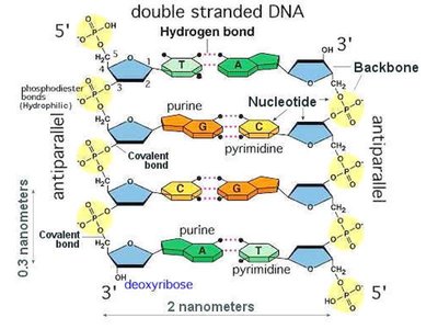
Purines: Double-ring structures (Adenine and Guanine)
Pyrimidines: Single-ring structures (Cytosine, Thymine in DNA, Uracil in RNA)

Base Pairing Rules
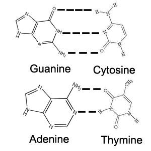
In DNA, specific base pairing occurs due to hydrogen bonding:
Adenine (A) pairs with Thymine (T) via 2 hydrogen bonds
Cytosine (C) pairs with Guanine (G) via 3 hydrogen bonds
In RNA, Uracil (U) replaces Thymine and pairs with Adenine.
Antiparallel Structure of DNA
The two strands of DNA run in opposite directions, described as antiparallel. One strand runs 5’ to 3’, and the other runs 3’ to 5’. This orientation is crucial for replication and function.
The 5’ end has a phosphate group attached to the 5’ carbon of the sugar.
The 3’ end has a hydroxyl group attached to the 3’ carbon of the sugar.
Summary Table: DNA vs. RNA
Characteristic | DNA | RNA |
|---|---|---|
Sugar | Deoxyribose | Ribose |
Bases | A, T, C, G | A, U, C, G |
Strands | Double | Single |
Function | Genetic information storage | Protein synthesis, gene regulation |
Applications and Activities
Artificial Selection Lab
Artificial selection is a process by which humans select for desirable traits in organisms. In the lab, students plant and observe the next generation of plants, following a protocol to ensure consistency and accurate data collection. This process models how genetic information is passed and selected across generations.
DNA Modeling Activities
Hands-on activities, such as using your body to represent the components of DNA (sugar, phosphate, base), help reinforce the structure and function of nucleic acids. These activities illustrate the double helix, base pairing, and antiparallel nature of DNA.
Key Terms and Concepts
Denaturation: Loss of protein structure due to environmental changes.
Nucleotide: Monomer of nucleic acids, composed of a sugar, phosphate, and nitrogenous base.
Purine: Double-ring nitrogenous base (Adenine, Guanine).
Pyrimidine: Single-ring nitrogenous base (Cytosine, Thymine, Uracil).
Antiparallel: Orientation of DNA strands running in opposite directions.
Equations and Chemical Structures
General nucleotide structure:
Base pairing:
Additional info: The notes above expand on the original content by providing definitions, explanations of key terms, and context for laboratory and classroom activities. Tables and images are included only where they directly reinforce the explanation of nucleic acid structure and function.
