 Back
Proteins and Nucleic Acids: Structure, Function, and Biological Importance
Study Guide - Smart Notes
 Tailored notes based on your materials, expanded with key definitions, examples, and context.
Proteins and Nucleic Acids
Overview of Biomolecules
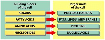
Biomolecules are essential components of living cells, serving as both structural and functional units. The four major types of biomolecules are carbohydrates, lipids, proteins, and nucleic acids. Each is built from specific monomers and forms larger macromolecular structures.
Carbohydrates: Built from sugars, form polysaccharides.
Lipids: Built from fatty acids, form fats, lipids, and membranes.
Proteins: Built from amino acids, form functional proteins.
Nucleic Acids: Built from nucleotides, form DNA and RNA.
Proteins
Amino Acids: The Building Blocks of Proteins
Amino acids are organic molecules composed of carbon (C), hydrogen (H), oxygen (O), nitrogen (N), and sometimes sulfur (S). They are the monomers that polymerize to form polypeptides and ultimately functional proteins.
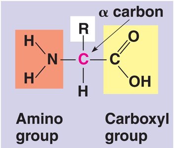
Structure: Each amino acid contains an amino group (-NH2), a carboxyl group (-COOH), a hydrogen atom, and a unique R group attached to a central α carbon.
R Group: The R group determines the chemical properties and reactivity of the amino acid.
Isoforms: All amino acids except glycine exist in D- and L- isoforms; only L-isoforms are found in proteins.
Peptide Bonds and Polypeptides
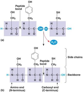
Amino acids are linked together by peptide bonds, which are covalent bonds formed between the carboxyl group of one amino acid and the amino group of another. This process occurs via a dehydration reaction, releasing water (H2O).
Peptide Bond: The bond joining two amino acids in a polypeptide chain.
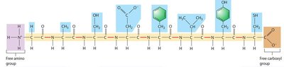
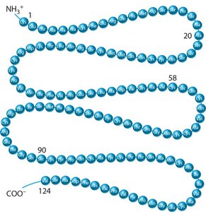
Polypeptide: A linear sequence of amino acids joined by peptide bonds; conventionally, chains longer than 10 amino acids are called polypeptides.
N-terminus and C-terminus: The ends of a polypeptide chain, with the N-terminus (amino end) and C-terminus (carboxyl end).
Protein Structure: Hierarchy and Function
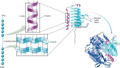
Proteins exhibit a hierarchical structure, with each level contributing to the overall shape and function. The four levels are primary, secondary, tertiary, and quaternary structure.
Primary Structure: The linear sequence of amino acids, determined by genetic information.
Secondary Structure: Local folding patterns such as α helices and β sheets, stabilized by hydrogen bonds.
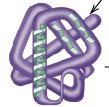
Tertiary Structure: The overall three-dimensional shape of a single polypeptide, determined by interactions among side chains (R groups).
Quaternary Structure: The assembly of two or more polypeptides into a functional protein complex.
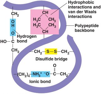
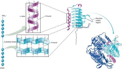
Types of Bonds in Protein Structure
Hydrogen Bonds: Weak individually, but strong collectively; stabilize secondary and tertiary structures.
Ionic Bonds: Form between oppositely charged side chains.
Hydrophobic Interactions: Nonpolar side chains cluster away from water.
Van der Waals Forces: Weak interactions between atoms in close proximity.
Disulfide Bridges: Covalent bonds between cysteine residues, stabilizing protein structure.
Protein Conformation and Denaturation
The specific three-dimensional shape (conformation) of a protein is critical to its function. Environmental factors such as temperature, pH, and salt concentration can disrupt these bonds, leading to denaturation and loss of function. Some proteins can be renatured with the help of chaperone proteins.
Denaturation: Loss of proper folding and function due to environmental changes.
Chaperone Proteins: Assist in folding and refolding of polypeptides.
Protein Domains
Domains are modular units within a protein that have distinct structures and functions. Different proteins may share similar domains, contributing to similar functions.
Functions of Proteins
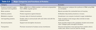
Proteins are involved in a wide range of cellular functions, including gene expression, movement, defense, catalysis, signaling, structure, and transport.
Category | Function | Examples |
|---|---|---|
Proteins involved in gene expression and regulation | Make mRNA from DNA template; synthesize polypeptides from mRNA; regulate genes | RNA polymerase catalyzes synthesis of RNA using DNA as a template |
Motor proteins | Initiate movement | Myosin provides contractile force of muscles |
Defense proteins | Protect organisms against disease | Antibodies help destroy bacteria or viruses |
Metabolic enzymes | Increase rates of chemical reactions | Hexokinase is an enzyme involved in sugar metabolism |
Cell-signaling proteins | Enable cells to communicate with each other and with their environment | Insulin regulates sugar levels in blood |
Structural proteins | Support and strengthen structures | Actin provides shape to cytoplasm of plant and animal cells; Collagen gives strength to skin |
Transporters | Promote movement of solutes across membranes | Glucose transporters move glucose from outside cells to inside cells; glucose can be used for energy |
Nucleic Acids
Nucleotides: The Building Blocks of Nucleic Acids
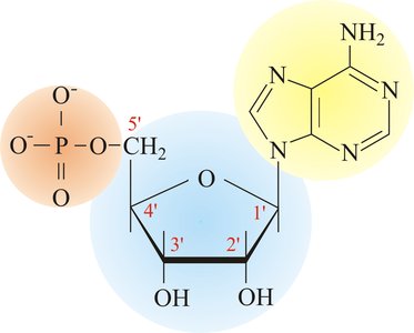
Nucleic acids are polymers of nucleotides, which are composed of carbon (C), hydrogen (H), oxygen (O), nitrogen (N), and phosphorus (P). Nucleotides consist of three parts: a phosphate group, a pentose sugar, and a nitrogen-containing base.
Phosphate Group: Provides negative charge and links nucleotides together.
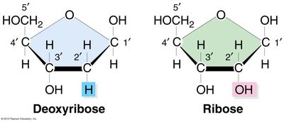
Pentose Sugar: Either ribose (in RNA) or deoxyribose (in DNA).
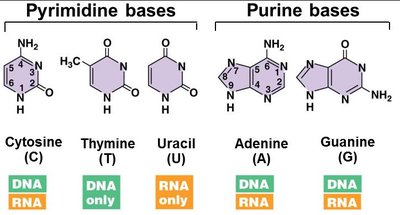
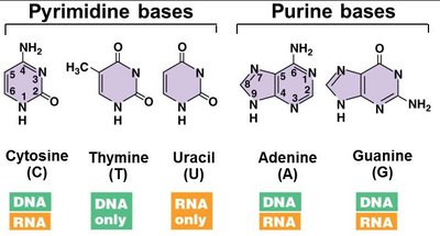
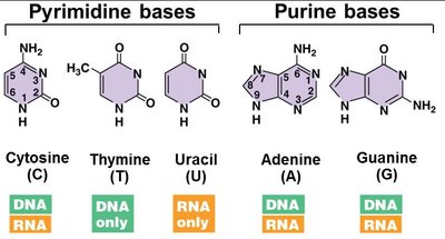
Nitrogen Base: Purines (adenine, guanine) and pyrimidines (cytosine, thymine, uracil).
Types of Nucleic Acids: DNA and RNA
DNA (deoxyribonucleic acid) stores genetic information, while RNA (ribonucleic acid) is transcribed from DNA and used to express genetic information as proteins.
DNA: Double-stranded, contains adenine (A), thymine (T), guanine (G), and cytosine (C).
RNA: Single-stranded, contains adenine (A), uracil (U), guanine (G), and cytosine (C).
Base Pairing in DNA and RNA
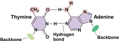
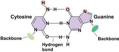
In DNA, nitrogen bases pair via hydrogen bonds: adenine pairs with thymine (A=T), and guanine pairs with cytosine (G≡C). In RNA, uracil replaces thymine.
Base Pairing:
Hydrogen Bonds: Hold the two strands of DNA together; these bonds are relatively weak compared to covalent bonds.
Structure of DNA
Nucleotides in a single DNA strand are linked by phosphodiester bonds, forming a sugar-phosphate backbone. Double-stranded DNA consists of two antiparallel strands held together by hydrogen bonds between complementary bases.
Phosphodiester Bond: Covalent bond joining the 5' phosphate group of one nucleotide to the 3' hydroxyl group of another.
Antiparallel Strands: The two DNA strands run in opposite directions.
Summary Table: Comparison of Proteins and Nucleic Acids
Type | Structure | Key Functions | Examples |
|---|---|---|---|
Proteins | Polypeptide is a structural unit composed of a linear sequence of amino acids. A protein is a functional unit composed of one or more polypeptides. | Proteins play a key role in cell structure and carry out a diverse array of cellular functions. For example, there are proteins involved with gene expression and regulation, motor proteins, defense proteins, cell-signaling proteins, metabolic enzymes, structural proteins, and transporters. | See Table 3.3 |
Nucleic acids | A nucleic acid is a linear sequence of nucleotides; DNA is double-stranded. | DNA stores genetic information in units called genes. RNA is made from DNA and provides access to that information. | DNA and RNA |




Example: Hemoglobin is a protein composed of four polypeptide subunits, each with a specific tertiary structure, forming a functional quaternary structure. DNA is a double-stranded nucleic acid that stores genetic information, while RNA is single-stranded and involved in protein synthesis.
Additional info: The notes expand on brief points by providing definitions, examples, and academic context for each topic, ensuring completeness and clarity for exam preparation.
