 Back
Bio 100 LEC Chapter 12 Module 3
Study Guide - Smart Notes
 Tailored notes based on your materials, expanded with key definitions, examples, and context.
Regulation of the Eukaryotic Cell Cycle
Overview of the Eukaryotic Cell Cycle
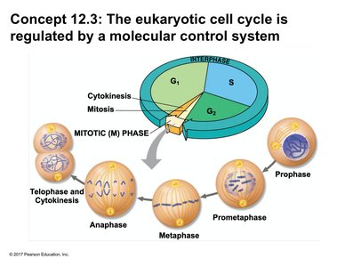
The eukaryotic cell cycle is a highly regulated process that ensures the accurate duplication and division of the cell's genetic material. The cell cycle consists of interphase (G1, S, and G2 phases) and the mitotic (M) phase, which includes mitosis and cytokinesis. Proper regulation is essential for maintaining genomic integrity and normal cellular function.
Interphase: The cell grows (G1), replicates its DNA (S), and prepares for division (G2).
M Phase: The cell undergoes mitosis (nuclear division) and cytokinesis (cytoplasmic division).
Regulation: Molecular control systems ensure that each phase is completed before the next begins.
Cell Cycle Checkpoints and Control Mechanisms
Major Cell Cycle Checkpoints
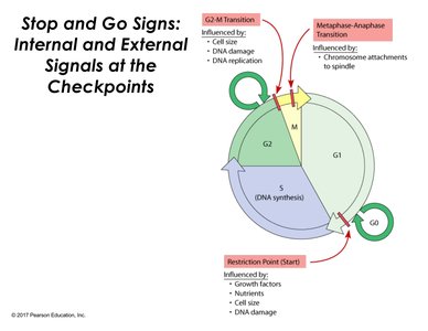
Checkpoints are critical control points where the cell assesses whether to proceed with division. These checkpoints help maintain the fidelity of cell division by monitoring internal and external conditions.
Restriction Point (G1 to S): Influenced by growth factors, nutrient availability, cell size, and DNA integrity. Cells that do not pass this checkpoint may enter a quiescent state (G0).
G2 to M Transition: Ensures the cell is the correct size, has undamaged DNA, and has completed DNA replication.
Metaphase-Anaphase Transition: Checks that all chromosomes are properly attached to the spindle apparatus before separation.
G0 Phase: Cellular Quiescence
The G0 phase is a non-dividing state that cells may enter from G1 if conditions are not favorable for division. Cells can remain in G0 temporarily or permanently, depending on their type and function.
Quiescent State: Cells are metabolically active but not actively preparing to divide.
Reversibility: Some cells can re-enter the cell cycle from G0 in response to appropriate signals.
Molecular Control of the Cell Cycle
Cytoplasmic Regulation of Cell Cycle Progression
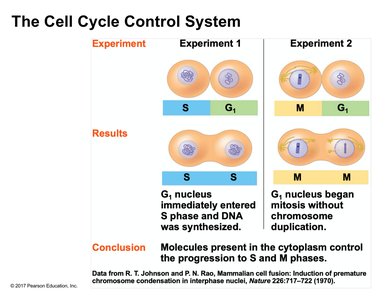
Experiments involving the fusion of cells at different cell cycle stages demonstrate that molecules in the cytoplasm can trigger progression through the cell cycle. These molecules are not confined to the nucleus but are present in the shared cytoplasm of fused cells (heterokaryons).
Key Finding: S phase cytoplasm can induce DNA synthesis in G1 nuclei; M phase cytoplasm can induce mitosis in G1 nuclei without DNA replication.
Conclusion: Cytoplasmic factors regulate entry into S and M phases.
Cyclins and Cyclin-Dependent Kinases (CDKs)
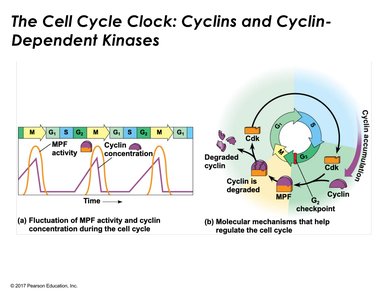
The cell cycle is regulated by cyclin-dependent kinases (CDKs), which are enzymes that require binding to cyclins for activation. The concentration of cyclins fluctuates throughout the cell cycle, controlling the activity of CDKs and thus the progression through different phases.
CDKs: Enzymes present throughout the cell cycle but active only when bound to cyclins.
Cyclins: Regulatory proteins whose levels rise and fall during the cell cycle.
MPF (Maturation-Promoting Factor): A complex of cyclin and CDK that triggers entry into mitosis.
Regulation: Cyclin accumulation and degradation control CDK activity and cell cycle transitions.
Mechanism of CDK Activation and Protein Phosphorylation
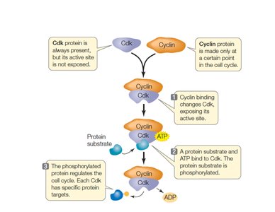
CDKs are inactive unless bound to cyclins. Upon binding, the CDK's active site is exposed, allowing it to phosphorylate target proteins using ATP. Phosphorylation of these proteins regulates various cell cycle events, such as spindle assembly and chromosome separation.
Inactive CDK: Active site is blocked until cyclin binds.
Activation: Cyclin binding exposes the active site, enabling kinase activity.
Phosphorylation: CDK transfers a phosphate group from ATP to a protein substrate, often activating the substrate.
External Regulation of the Cell Cycle
Growth Factors and Cell Division
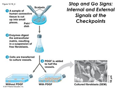
External signals, such as growth factors, can stimulate cell division. Platelet-derived growth factor (PDGF) is an example of a growth factor that promotes the proliferation of fibroblasts in vitro. Cells require both a suitable substrate and appropriate external signals to divide.
Growth Factors: Proteins that stimulate cell growth and division.
Mitogens: Substances that specifically trigger mitosis.
Experimental Evidence: Cells cultured with PDGF show increased proliferation compared to those without PDGF.
Anchorage Dependence and Density-Dependent Inhibition
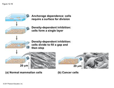
Normal mammalian cells exhibit anchorage dependence (requiring a surface for division) and density-dependent inhibition (stopping division when the culture surface is fully occupied). These mechanisms prevent uncontrolled cell proliferation.
Anchorage Dependence: Cells must be attached to a substrate to divide.
Density-Dependent Inhibition: Cells stop dividing when they form a single layer and fill available space (100% confluency).
Cell Communication: Cells sense crowding and inhibit further division until space becomes available.
Loss of Cell Cycle Control and Cancer
Characteristics of Cancer Cells
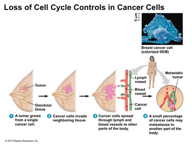
Cancer cells evade normal regulatory mechanisms, leading to uncontrolled proliferation. They do not exhibit anchorage dependence or density-dependent inhibition, allowing them to grow without attachment and to form multiple layers in culture. This loss of regulation underlies tumor formation and metastasis.
Tumor Formation: Cancer cells proliferate uncontrollably, forming masses (tumors).
Invasion: Cancer cells invade neighboring tissues and can enter blood or lymphatic vessels.
Metastasis: The spread of cancer cells to distant sites, forming secondary tumors.
Experimental Analysis: Cell Cycle Inhibitors in Cancer Treatment
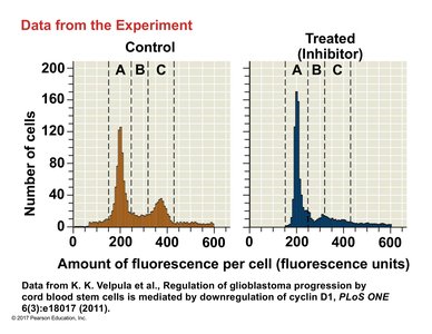
Flow cytometry can be used to analyze the effects of cell cycle inhibitors on cancer cells. By staining DNA with a fluorescent dye, researchers can determine the distribution of cells across different cell cycle phases. Inhibitors can arrest cells at specific checkpoints, reducing proliferation and offering potential therapeutic benefits.
Control vs. Treated: Inhibitor-treated cells show altered cell cycle distribution compared to controls, often accumulating in a specific phase.
Application: Understanding cell cycle regulation aids in developing targeted cancer therapies.
Summary Table: Key Cell Cycle Regulatory Mechanisms
Checkpoint | Main Regulatory Factors | Outcome if Not Passed |
|---|---|---|
G1 (Restriction Point) | Growth factors, nutrients, cell size, DNA damage | Entry into G0 (quiescence) |
G2-M Transition | Cell size, DNA damage, DNA replication | Cell cycle arrest in G2 |
Metaphase-Anaphase | Chromosome attachment to spindle | Cell cycle arrest in metaphase |
Key Terms and Concepts
Cyclin: Regulatory protein whose concentration fluctuates during the cell cycle.
Cyclin-Dependent Kinase (CDK): Enzyme that, when activated by cyclin, phosphorylates target proteins to regulate the cell cycle.
MPF (Maturation-Promoting Factor): Cyclin-CDK complex that triggers mitosis.
Growth Factor: External signal that stimulates cell growth and division.
Anchorage Dependence: Requirement for a surface to divide.
Density-Dependent Inhibition: Inhibition of cell division when cells are crowded.
Metastasis: Spread of cancer cells to distant tissues.
Relevant Equations
Phosphorylation Reaction Catalyzed by CDK:
