 Back
lecture 4 Respiration and Excretion: Mechanisms and Adaptations in Animals
Study Guide - Smart Notes
 Tailored notes based on your materials, expanded with key definitions, examples, and context.
lecture 4 Respiratory Pigments and Oxygen Transport
Hemoglobin and Other Respiratory Pigments
Respiratory pigments are specialized proteins that significantly enhance the oxygen-carrying capacity of blood in animals. These pigments are crucial for efficient oxygen transport and delivery to tissues.
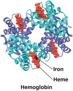
Hemoglobin (Hb): The primary respiratory pigment in vertebrates and some invertebrates. Each hemoglobin molecule consists of four subunits (two alpha and two beta chains), each containing an iron-bearing heme group capable of binding one O2 molecule.
Hemocyanin: Found in arthropods and many molluscs, uses copper instead of iron as the oxygen-binding component.
Oxygen Binding: Each hemoglobin molecule can carry up to four O2 molecules, greatly increasing the oxygen content of blood compared to plasma alone.
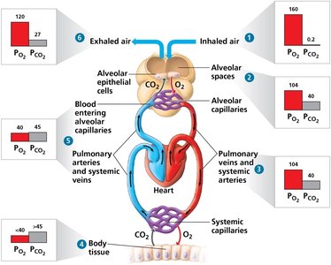
Loading and Unloading of O2 and CO2
Oxygen and carbon dioxide are exchanged between the environment and animal tissues via diffusion, following their respective partial pressure gradients.
O2 Diffusion: Moves from areas of high partial pressure (alveolar spaces) to low partial pressure (lung capillaries and then tissues).
CO2 Diffusion: Moves from tissues (high PCO2) to systemic capillaries, then to alveolar spaces for exhalation.
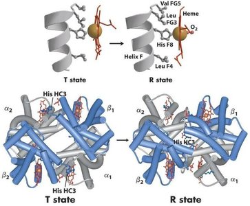
Tense and Relaxed States of Hemoglobin
Hemoglobin exhibits two major conformational states that influence its affinity for oxygen:
Tense (T) State: Stabilized by ionic bonds and salt bridges; Fe2+ is slightly out of the plane of the heme, resulting in low oxygen affinity and low cooperativity.
Relaxed (R) State: High affinity for oxygen; binding of one O2 increases the likelihood of additional O2 binding (high cooperativity).
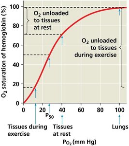
Hemoglobin-Oxygen Dissociation Curve
The relationship between the partial pressure of oxygen (PO2) and hemoglobin saturation is sigmoidal, reflecting cooperative binding. The P50 value indicates the partial pressure at which hemoglobin is 50% saturated with oxygen.
Cooperativity: Small changes in PO2 result in large changes in O2 saturation, facilitating efficient oxygen loading and unloading.
The Bohr Shift
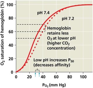
The Bohr effect describes how increased CO2 production during cellular respiration lowers blood pH, decreasing hemoglobin's affinity for O2 and promoting oxygen release to tissues.
Mechanism: Lower pH (higher CO2) increases P50, shifting the dissociation curve to the right.
BPG (2,3-Bisphosphoglycerate) Regulation
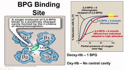
BPG binds to deoxygenated hemoglobin, stabilizing the T state and reducing oxygen affinity, which facilitates oxygen release in tissues, especially under conditions such as high altitude.
Myoglobin
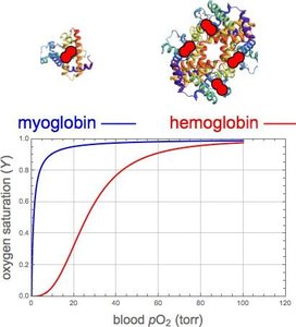
Myoglobin is a monomeric oxygen-binding protein found in muscle tissue. It has a higher affinity for oxygen than hemoglobin and serves as an emergency oxygen reserve.
No Cooperativity: Myoglobin binds oxygen independently, unlike hemoglobin.
CO2 Transport
Hemoglobin also plays a role in transporting carbon dioxide and buffering blood pH.
~7% of CO2 is dissolved in plasma.
~23% binds to hemoglobin (carbaminohemoglobin).
~70% is transported as bicarbonate ions (HCO3-).
CO Poisoning
Carbon monoxide (CO) binds to hemoglobin with much higher affinity than oxygen, forming carboxyhemoglobin (HbCO) and preventing oxygen transport, which can be fatal.
Neural Control of Breathing
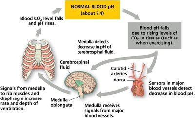
The medulla oblongata in the brain is the primary control center for breathing, responding to changes in blood CO2 and O2 levels detected by sensors in the aorta and carotid arteries.
Respiratory Adaptations of Diving Mammals
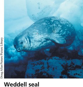
Diving mammals, such as Weddell seals, have evolved adaptations for extended underwater activity, including high blood volume, increased myoglobin in muscles, and the ability to reduce oxygen consumption by selective blood flow and anaerobic metabolism.
Ice Fish Adaptations
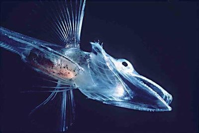
Ice fish lack hemoglobin but survive in cold, oxygen-rich waters due to high body fluid concentrations, large hearts, and extensive capillary networks.
Osmoregulation and Excretion
Principles of Osmoregulation
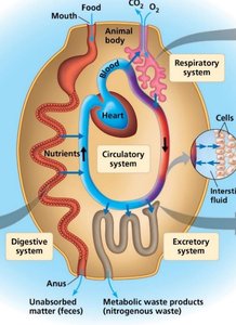
Osmoregulation is the process by which animals regulate the balance of water and solutes in their bodies, essential for maintaining cellular function and homeostasis.
Osmolarity: Total concentration of osmotically active particles per liter of solution (Osm/L).
Osmolality: Total concentration per kilogram of solvent (Osm/kg).
Animals face different challenges depending on their environment (marine, freshwater, terrestrial).
Osmosis, Osmolarity, and Osmolality
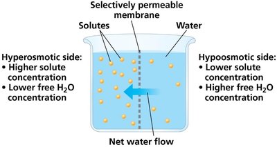
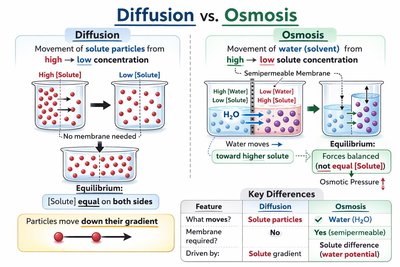
Osmosis is the passive movement of water across a selectively permeable membrane, driven by differences in solute concentration (osmolarity/osmolality).
Hyperosmotic: Higher solute concentration, lower free water.
Hypoosmotic: Lower solute concentration, higher free water.
Isoosmotic: Equal solute concentrations on both sides.
Aquaporins
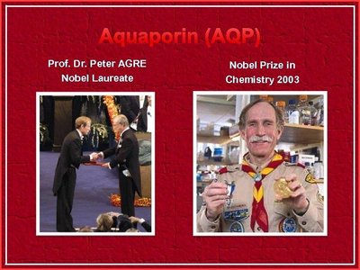
Aquaporins are membrane proteins that facilitate rapid water transport across cell membranes, crucial for osmoregulation in many tissues.
Discovery of aquaporins revolutionized understanding of water movement in cells.
Osmoregulatory Strategies
Animals employ different strategies to cope with osmotic challenges:
Osmoconformers: Maintain internal osmolarity equal to their environment (e.g., most marine invertebrates).
Osmoregulators: Regulate internal osmolarity, often expending energy to do so (e.g., marine vertebrates, freshwater animals).
Stenohaline: Tolerate only narrow changes in external osmolarity.
Euryhaline: Can survive large fluctuations in external osmolarity (e.g., salmon).
Osmoregulation in Aquatic and Terrestrial Animals
Marine Fish: Hypoosmotic to seawater, lose water by osmosis, drink seawater, excrete salts.
Freshwater Fish: Hyperosmotic to environment, gain water by osmosis, excrete dilute urine, actively uptake salts.
Terrestrial Animals: Adaptations to reduce water loss include waterproof body coverings, nocturnal behavior, and metabolic water production.
Transport Epithelia in Osmoregulation
Specialized epithelial cells actively transport solutes for osmoregulation, such as the nasal salt glands in marine birds.
Forms of Nitrogenous Wastes
Animals excrete nitrogenous wastes in different forms, depending on their evolutionary history and habitat:
Ammonia: Highly toxic, requires large amounts of water for excretion (common in aquatic animals).
Urea: Less toxic, conserves water, requires energy to synthesize (mammals, amphibians, some fish).
Uric Acid: Least toxic, excreted as a paste, conserves water, energetically expensive (insects, birds, reptiles).
Excretory Systems and Processes
Excretory systems perform four main functions:
Filtration: Filtering of body fluids.
Reabsorption: Reclaiming valuable solutes.
Secretion: Adding nonessential solutes and wastes to the filtrate.
Excretion: Removal of processed filtrate containing nitrogenous wastes.
Types of Excretory Organs
Protonephridia: Network of dead-end tubules (e.g., flatworms).
Metanephridia: Tubules in each segment (e.g., earthworms).
Malpighian Tubules: Remove wastes from hemolymph (e.g., insects).
Kidneys: Complex organs in vertebrates, filter blood and produce urine.
The Mammalian Kidney and Nephron Function
The nephron is the functional unit of the kidney, responsible for filtering blood and forming urine through a series of specialized segments:
Proximal Tubule: Reabsorbs ions, water, and nutrients; secretes toxins.
Loop of Henle: Descending limb reabsorbs water; ascending limb reabsorbs salt but not water.
Distal Tubule and Collecting Duct: Further regulate ion and water balance; collecting duct concentrates urine.
Countercurrent Multiplier System
The loop of Henle creates an osmotic gradient in the renal medulla, allowing the kidney to produce urine that is hyperosmotic to blood.
Hormonal Regulation of Kidney Function
Antidiuretic Hormone (ADH): Increases water reabsorption in the collecting duct by promoting aquaporin insertion, reducing urine volume and lowering blood osmolarity.
Renin-Angiotensin-Aldosterone System (RAAS): Responds to decreased blood pressure by increasing sodium and water reabsorption, raising blood volume and pressure.
Atrial Natriuretic Peptide (ANP): Opposes RAAS and ADH, reducing blood volume when it is too high.
Solute Gradients and Water Conservation
The ability to produce hyperosmotic urine is a key adaptation for terrestrial life, relying on energy-dependent solute transport and the countercurrent multiplier system.
The Two-Solute Model
NaCl and urea are the primary solutes establishing the osmotic gradient in the kidney, enabling efficient water reabsorption and urine concentration.
