 Back
BackSensory and Motor Mechanisms: Muscle, Skeletal Systems, and Locomotion
Study Guide - Smart Notes
 Tailored notes based on your materials, expanded with key definitions, examples, and context.
Sensory and Motor Mechanisms
Introduction
Sensory and motor mechanisms are essential for animal movement and interaction with the environment. This section explores the structure and function of muscles, skeletal systems, and the principles of locomotion in animals.
Muscle Function and Structure
Muscle Contraction: Overview
Muscle contraction is a physiological process that enables movement in animals. It is initiated by signals from the nervous system and involves the interaction of protein filaments within muscle cells.
Muscle activity is triggered by input from motor neurons.
Relies on thin filaments (actin) and thick filaments (myosin).
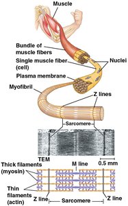
Muscle fibers are organized into bundles, each containing myofibrils composed of repeating units called sarcomeres.
Sarcomeres are the contractile units of skeletal muscle, bordered by Z lines where thin filaments attach.
Sliding Filament Model of Muscle Contraction
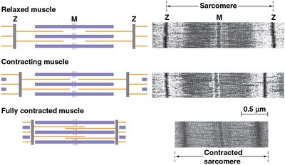
The sliding filament model explains how muscles contract at the molecular level. During contraction, thin filaments slide past thick filaments, shortening the sarcomere and thus the muscle.
In a relaxed muscle, sarcomeres are at their maximum length.
During contraction, the overlap between actin and myosin increases, shortening the sarcomere.
Muscle contraction is powered by ATP hydrolysis.
Molecular Mechanism of Contraction
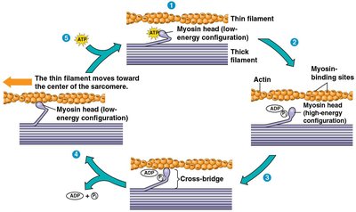
Muscle contraction is regulated by the exposure of myosin-binding sites on actin filaments, which is controlled by calcium ions and regulatory proteins.
At rest, proteins bound to actin prevent myosin from binding.
Motor neurons release acetylcholine, triggering an action potential and the release of calcium ions from the sarcoplasmic reticulum.
Calcium binds to regulatory proteins, exposing myosin-binding sites on actin.
Myosin heads bind to actin, forming cross-bridges and pulling the thin filaments toward the center of the sarcomere.
ATP is required for myosin head detachment and re-cocking.
Regulation of Muscle Contraction Strength
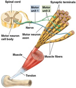
The strength of a muscle contraction depends on the number of muscle fibers activated and the frequency of stimulation.
Motor unit: A single motor neuron and all the muscle fibers it innervates.
Recruitment of more motor units increases contraction strength.
Higher stimulation rates lead to stronger, sustained contractions (tetanus).
Skeletal Systems
Types of Skeletal Systems
Skeletal systems provide support, protection, and leverage for movement. There are three main types:
Hydrostatic skeleton: Fluid-filled compartments provide support (e.g., cnidarians, annelids).
Exoskeleton: Hard external covering (e.g., arthropods, molluscs).
Endoskeleton: Internal skeleton made of bone and/or cartilage (e.g., vertebrates, echinoderms).
Hydrostatic Skeleton
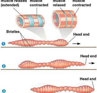
Hydrostatic skeletons rely on fluid pressure within a closed body compartment. Muscles contract against the fluid, changing the animal's shape and enabling movement such as peristalsis.
Exoskeleton
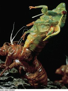
Exoskeletons are rigid structures that cover the body surface. They provide protection and points of muscle attachment but must be shed for growth (molting).
Arthropod exoskeletons are made of chitin and are jointed for flexibility.
Endoskeleton
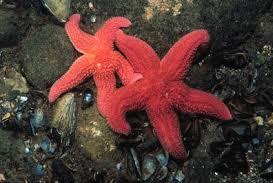
Endoskeletons are internal frameworks that can grow with the organism. They are found in echinoderms and chordates and are composed of bone, cartilage, or both.
Human Skeletal System
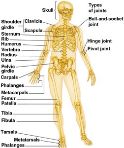
The human skeleton is divided into the axial and appendicular skeletons and contains various types of joints for movement.
Axial skeleton: Skull, vertebral column, and rib cage.
Appendicular skeleton: Limbs and girdles.
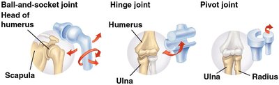
Joints
Joints are the sites where two or more bones meet, allowing for different types of movement.
Ball-and-socket joints: Allow rotation and movement in all directions (e.g., shoulder, hip).
Hinge joints: Permit movement in one plane (e.g., knee, elbow).
Pivot joints: Allow rotational movement (e.g., between radius and ulna).
Locomotion
Principles of Locomotion
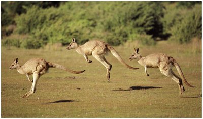
Locomotion is the ability of an organism to move from one place to another. Different environments require specialized adaptations for movement.
Swimming: Movement through water, often using fins or undulating body movements.
Land movement: Includes walking, crawling, and running, requiring support against gravity.
Flying: Movement through the air, requiring adaptations such as wings and lightweight bodies.
Summary Table: Types of Skeletal Systems
Type | Main Features | Examples |
|---|---|---|
Hydrostatic | Fluid-filled compartments, flexible, support via pressure | Cnidarians, annelids |
Exoskeleton | External, rigid, must be molted for growth | Arthropods, molluscs |
Endoskeleton | Internal, can grow, made of bone/cartilage | Vertebrates, echinoderms |
Key Terms
Sarcomere: The basic contractile unit of muscle fiber.
Actin: Protein forming thin filaments in muscle.
Myosin: Protein forming thick filaments in muscle.
Motor unit: A motor neuron and all the muscle fibers it controls.
Exoskeleton: Hard external skeleton.
Endoskeleton: Internal skeleton.
Hydrostatic skeleton: Support system using fluid pressure.
Equations
Muscle contraction and force generation can be described by the following relationship:
Where is force, is mass, and is acceleration.
ATP hydrolysis during muscle contraction:
