Back
BackSensory and Motor Mechanisms: Structure and Function in Animal Physiology
Study Guide - Smart Notes
 Tailored notes based on your materials, expanded with key definitions, examples, and context.
Sensory and Motor Mechanisms
Introduction
Sensory and motor mechanisms are essential for animals to detect environmental changes and respond appropriately. These processes involve specialized cells and organs that convert various forms of energy into signals, which are then interpreted by the nervous system to produce coordinated motor outputs.
Concept 50.1: Sensory Receptors and Signal Transduction
Sensory Reception and Transduction
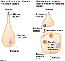
Sensory reception is the detection of stimuli by sensory receptors, which may be neurons or non-neuronal cells. These receptors interact with both internal and external stimuli, converting stimulus energy into changes in membrane potential, a process known as sensory transduction. The resulting change in membrane potential is called a receptor potential, which is a graded potential whose magnitude varies with stimulus strength.
Transmission of Sensory Information
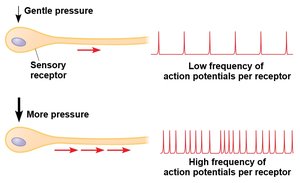
Sensory information is transmitted through the nervous system as action potentials. The frequency of action potentials generated by a sensory neuron is proportional to the intensity of the stimulus. In neurons that spontaneously generate action potentials, a stimulus alters the rate of firing.
Perception
Perception is the brain’s interpretation of sensory stimuli. Although all action potentials are similar, the brain distinguishes the type and location of a stimulus based on the neural pathway the signal travels. This allows for the differentiation of sensory modalities such as touch, sound, and light.
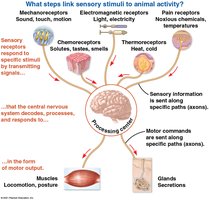
Types of Sensory Receptors
Mechanoreceptors: Detect physical deformation (e.g., touch, pressure, sound).
Chemoreceptors: Respond to chemical stimuli (e.g., taste, smell).
Electromagnetic receptors: Detect light, electricity, and magnetism.
Thermoreceptors: Sense temperature changes.
Pain receptors (nociceptors): Detect harmful conditions such as extreme heat, pressure, or chemicals.
Concept 50.2: Mechanoreceptors in Hearing and Equilibrium
Mechanoreceptors and Body Position
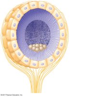
Mechanoreceptors are crucial for detecting movement and position. In invertebrates, organs called statocysts contain mechanoreceptors that sense the movement of granules called statoliths, providing information about gravity and orientation.
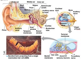
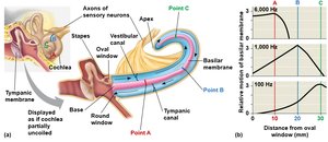
Hearing in Insects and Humans
Many insects detect sound through body hairs or tympanic membranes. In humans, the ear transduces sound waves into nerve impulses using hair cells with hairlike projections. The cochlea distinguishes pitch due to the varying properties of the basilar membrane along its length.
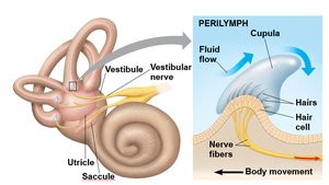
Equilibrium in Vertebrates
The inner ear contains the utricle, saccule, and semicircular canals, which detect linear and angular movements. Hair cells in these structures respond to fluid movement, allowing the brain to perceive balance and orientation.
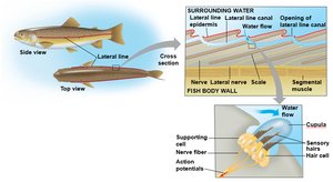
Lateral Line System in Aquatic Vertebrates
Fishes and aquatic amphibians possess a lateral line system with mechanoreceptors that detect water movement, aiding in navigation and prey detection.
Concept 50.3: Visual Receptors and Light Detection
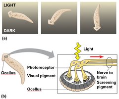
Light-Detecting Organs
Animals possess a variety of light-detecting organs, from simple eyespots in planarians to complex compound and single-lens eyes. All light detectors contain photoreceptors with light-absorbing pigments.
Compound and Single-Lens Eyes
Compound eyes, found in insects and crustaceans, consist of many ommatidia and are highly sensitive to movement. Single-lens eyes, present in vertebrates and some invertebrates, function like a camera, focusing light onto a retina.
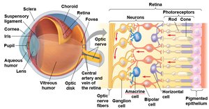
The Vertebrate Visual System
The vertebrate eye contains layers including the sclera, choroid, retina, and lens. Light passes through the lens and strikes the retina, where rods and cones transduce light into neural signals. Rods are sensitive to low light, while cones enable color vision.
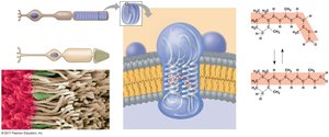
Visual Pigments and Phototransduction
Visual pigments consist of retinal (a light-absorbing molecule) bound to opsin proteins. In rods, this complex is called rhodopsin. Light absorption causes retinal to change shape, initiating a signal transduction cascade that alters neurotransmitter release.
Processing Visual Information
In darkness, rods and cones release glutamate, which is reduced upon light exposure, altering the activity of bipolar cells and ultimately ganglion cells, whose axons form the optic nerve.

Color Vision
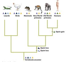
Color vision in humans is based on three types of cones, each with a distinct opsin sensitive to red, green, or blue light. Other vertebrates may have more or fewer types of cones, affecting their color perception.
Concept 50.4: Taste and Smell
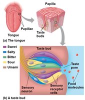
Taste (Gustation) in Mammals
There are five basic taste modalities: sweet, sour, salty, bitter, and umami. Taste receptor cells are organized into taste buds, primarily on the tongue. Each taste bud can detect all five tastes, and different receptor mechanisms are involved for each modality.
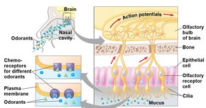
Smell (Olfaction) in Mammals
Olfactory receptor cells in the nasal cavity detect odorant molecules, triggering signal transduction pathways that generate action potentials. Mammals can distinguish thousands of odors, and taste and smell pathways interact in the brain.
Concept 50.5: Muscle Structure and Function
Muscle Types and Structure
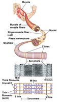
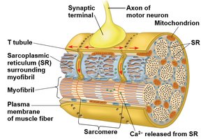
Muscle activity is based on the interaction of actin (thin) and myosin (thick) filaments. Vertebrate skeletal muscle is composed of bundles of fibers, each containing myofibrils organized into repeating units called sarcomeres. The regular arrangement of filaments gives skeletal muscle its striated appearance.
Sliding-Filament Model of Contraction
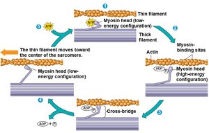
Muscle contraction occurs as myosin heads bind to actin, forming cross-bridges and pulling thin filaments toward the sarcomere center. This process is powered by ATP hydrolysis and involves repeated cycles of binding and release.
Role of Calcium and Regulatory Proteins
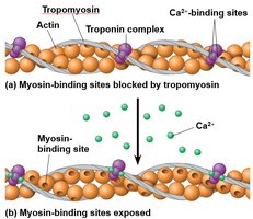
At rest, tropomyosin and the troponin complex block myosin-binding sites on actin. Calcium ions bind to troponin, causing a conformational change that exposes these sites, allowing contraction to occur. Muscle contraction is regulated by the concentration of Ca2+ in the cytosol.
Excitation-Contraction Coupling
An action potential in a motor neuron triggers acetylcholine release at the neuromuscular junction, depolarizing the muscle fiber membrane. The action potential travels along T tubules, causing the sarcoplasmic reticulum to release Ca2+, initiating contraction.
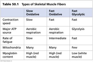
Muscle Fiber Types
Skeletal muscle fibers are classified based on contraction speed and metabolic pathway:
Type | Contraction Speed | Major ATP Source | Rate of Fatigue | Mitochondria | Myoglobin Content |
|---|---|---|---|---|---|
Slow Oxidative | Slow | Aerobic respiration | Slow | Many | High (red muscle) |
Fast Oxidative | Fast | Aerobic respiration | Intermediate | Many | High (red muscle) |
Fast Glycolytic | Fast | Glycolysis | Fast | Few | Low (white muscle) |
Other Muscle Types
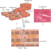
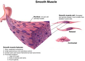
Cardiac muscle, found only in the heart, is striated and connected by intercalated disks, allowing coordinated contractions. Smooth muscle, found in the walls of hollow organs, is not striated and contracts more slowly, often under autonomic control.