 Back
Sensory and Motor Mechanisms: Structure, Function, and Integration
Study Guide - Smart Notes
 Tailored notes based on your materials, expanded with key definitions, examples, and context.
Sensory and Motor Mechanisms
Introduction to Sensory and Motor Systems
Sensory and motor mechanisms are essential for organisms to detect environmental changes and respond appropriately. Sensory receptors transduce various forms of energy into electrical signals, which are processed by the nervous system to generate perceptions and coordinate motor responses.
Sensory Reception and Transduction
Key Concepts in Sensory Reception
Sensation vs. Perception: Sensation is the process of detecting environmental stimuli, while perception is the brain's interpretation of these signals.
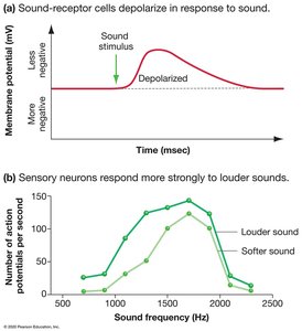
Sensory Transduction: The conversion of stimulus energy (e.g., light, sound, pressure) into a receptor potential, a graded change in membrane potential of the sensory receptor.
Receptor Potential: The magnitude of this potential varies with stimulus strength and can initiate action potentials in sensory neurons.
Transmission and Integration
Action potentials generated by sensory receptors are transmitted to the central nervous system (CNS).
Integration occurs in the CNS, where signals are processed and interpreted.
Perception depends on the neural pathway activated, not the nature of the stimulus itself.
Types of Sensory Receptors
Classification by Energy Transduced
Mechanoreceptors: Detect physical deformation (pressure, stretch, vibration, sound).
Chemoreceptors: Respond to chemical stimuli (solute concentration, specific molecules).
Electromagnetic Receptors: Detect light, electricity, and magnetism.
Thermoreceptors: Respond to temperature changes.
Pain Receptors (Nociceptors): Detect damaging or potentially damaging stimuli (excess heat, pressure, chemicals).
Mechanoreceptors in Human Skin
Mechanoreceptors are distributed throughout the skin and are responsible for sensing touch, pressure, vibration, and hair movement.
Chemoreceptors
Chemoreceptors can detect overall solute concentration or specific molecules. For example, osmoreceptors in the brain monitor blood osmolarity, while insect antennae detect pheromones.
Electromagnetic and Thermoreceptors
Electromagnetic receptors allow animals to sense light (vision), electric fields, and magnetic fields. Thermoreceptors help regulate body temperature by detecting heat or cold.
Pain Receptors
Pain receptors (nociceptors) are free nerve endings in the skin that respond to potentially harmful stimuli, triggering protective reflexes and pain perception.
Hearing and Equilibrium in Mammals
Mechanics of Hearing
The mammalian ear transduces sound waves into nerve impulses. The process involves the external ear collecting sound, the middle ear amplifying vibrations, and the inner ear (cochlea) converting these vibrations into electrical signals.
Pitch, Loudness, and Timbre
Pitch: Determined by the frequency of sound waves; different regions of the cochlea respond to different frequencies.
Loudness: Determined by the amplitude of sound waves; encoded by the frequency of action potentials.
Timbre: The quality of sound, determined by the complexity of the waveform.
Equilibrium
The vestibular system (utricle, saccule, and semicircular canals) detects head position, linear acceleration, and rotational movement, helping maintain balance.
Vertebrate Visual System
Structure and Function of the Eye
The vertebrate eye focuses light onto the retina, where photoreceptors (rods and cones) transduce light energy into electrical signals. The brain assembles these signals into visual images.
Photoreceptors: Rods and Cones
Rods: Highly sensitive to light, enable vision in dim conditions, do not distinguish color.
Cones: Less sensitive to light, responsible for color vision and visual acuity.
Phototransduction and Adaptation
Light absorption by photopigments (e.g., rhodopsin in rods) triggers a cascade that alters membrane potential and neurotransmitter release. Adaptation mechanisms allow the eye to adjust to varying light conditions.
Retinal Processing and Visual Pathways
Signals from photoreceptors are processed by bipolar, horizontal, amacrine, and ganglion cells in the retina. Lateral inhibition enhances image contrast. Ganglion cell axons form the optic nerve, transmitting visual information to the brain.
Color Vision and Visual Field
Color vision is based on three types of cones (red, green, blue), each with distinct absorption spectra.
The fovea is the region of highest visual acuity, densely packed with cones.
Chemical Senses: Taste and Smell
Gustation (Taste)
Taste buds on the tongue contain sensory receptor cells that detect tastants (chemicals responsible for sweet, salty, sour, bitter, and umami tastes).
Olfaction (Smell)
Olfactory receptors in the nasal cavity bind odorant molecules, initiating signal transduction pathways that result in action potentials sent to the olfactory bulb in the brain.
Muscle Structure and Function
Organization of Skeletal Muscle
Skeletal muscle fibers are composed of myofibrils, which contain repeating units called sarcomeres. Sarcomeres are made of thick (myosin) and thin (actin) filaments responsible for muscle contraction.
Sliding-Filament Model of Muscle Contraction
Muscle contraction occurs as myosin heads bind to actin, pulling thin filaments toward the center of the sarcomere. This process is powered by ATP hydrolysis.
Control of Muscle Tension
The nervous system regulates muscle contraction by varying the number of active motor units and the frequency of stimulation. Summation of twitches can lead to sustained contraction (tetanus).
Types of Skeletal Muscle Fibers
Oxidative fibers: Use aerobic respiration, rich in mitochondria and myoglobin, fatigue-resistant.
Glycolytic fibers: Use glycolysis, less myoglobin, fatigue more quickly.
Slow-twitch fibers: Contract slowly, sustain contractions, primarily oxidative.
Fast-twitch fibers: Contract rapidly, fatigue quickly, can be oxidative or glycolytic.
Other Muscle Types
Cardiac muscle: Found only in the heart, striated, electrically connected by intercalated disks, can generate action potentials without neural input.
Smooth muscle: Found in walls of hollow organs, non-striated, contractions are slow and can be initiated by the muscle itself or autonomic nervous system.
Skeletal Systems and Movement
Types of Skeletons
Endoskeleton: Internal skeleton made of bone and cartilage (e.g., mammals).
Exoskeleton: External skeleton (e.g., arthropods).
Hydrostatic skeleton: Fluid-filled cavity (e.g., annelids).
Antagonistic Muscle Arrangement
Skeletal muscles are arranged in antagonistic pairs (e.g., biceps and triceps) to enable movement in opposite directions. Movement is produced by contraction of one muscle and relaxation of its antagonist.
Types of Movable Joints
Ball-and-socket joint: Allows movement in multiple directions (e.g., shoulder).
Hinge joint: Allows movement in one plane (e.g., elbow).
Pivot joint: Allows rotational movement (e.g., neck).
Additional info: This guide integrates foundational concepts in sensory and motor biology, including receptor types, sensory pathways, muscle structure, and the mechanics of movement, as outlined in standard college-level biology curricula.
