 Back
BackSensory and Motor Mechanisms: Structure, Function, and Integration
Study Guide - Smart Notes
 Tailored notes based on your materials, expanded with key definitions, examples, and context.
Sensory and Motor Mechanisms
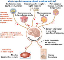
Overview of Sensory and Motor Pathways
Sensory and motor mechanisms are fundamental to animal physiology, enabling organisms to detect environmental stimuli and respond appropriately. Sensory receptors convert various forms of energy into electrical signals, which are processed by the nervous system to produce motor outputs.
Sensory receptors detect specific stimuli (light, sound, chemicals, temperature, pain, electric and magnetic fields).
Signal transduction converts stimulus energy into a cellular signal (action potential).
Central nervous system integrates sensory information and generates motor commands.
Motor output is executed by muscles (locomotion, posture) or glands (secretion).
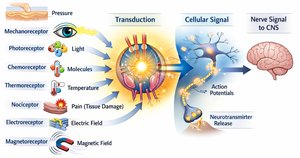
Types of Sensory Receptors
Sensory receptors are specialized to detect distinct forms of energy and transduce them into neural signals.
Mechanoreceptors: Detect pressure, stretch, vibration, and sound.
Photoreceptors: Respond to specific wavelengths of light.
Chemoreceptors: Sense molecules (solutes, tastes, smells).
Thermoreceptors: Detect temperature changes.
Nociceptors: Respond to harmful stimuli (pain, tissue damage).
Electroreceptors: Detect electric fields.
Magnetoreceptors: Sense magnetic fields.
Sensory Transduction and Neural Coding
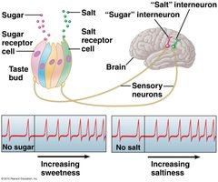
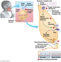
Taste Sensation and Neural Coding
Taste perception involves the detection of specific molecules by taste bud receptors, followed by signal transduction and neural coding.
Sweet taste: Sugar binds to sweet receptors, triggering a signaling pathway and receptor potential.
Transmission: Neurotransmitter release leads to action potentials in sensory neurons, which travel to the CNS.
Coding: Stronger stimuli produce higher action potential frequencies.
Perception: The brain interprets the activity in labeled pathways as specific taste qualities.
Receptor Types and Taste Perception
Different taste qualities are detected by distinct receptor mechanisms:
Salty: Na+ enters through channels, causing depolarization.
Sour: H+ (acids) cause depolarization.
Sweet: GPCRs (T1R receptors) mediate detection.
Umami: Glutamate receptor (e.g., MSG).
Bitter: Many GPCRs (40–80 genes), evolved to detect toxins.
Olfaction: Smell and Odorant Receptors
Olfactory System and Odor Coding
Olfactory receptor neurons detect odorants and transmit signals to the olfactory bulb, where odor identity is encoded by patterns of receptor activation.
Odorant binding: Initiates signal transduction and action potentials.
Olfactory receptor (OR) neurons: Each expresses one OR gene; neurons with the same OR project to the same glomerulus.
Odorant receptor gene family: Largest known gene family (~3–5% of human genome), encoding GPCRs with 7 transmembrane domains.
Gene number: Correlates with olfactory acuity; ~60% are pseudogenes in humans.
Auditory System: Hearing and Sound Transduction
Sound Waves and Hearing
Hearing involves the detection of sound waves, which are pressure changes traveling as sinusoidal waves.
Amplitude (dB): Determines loudness.
Frequency (Hz): Determines pitch.
Tonotopy: Spatial mapping of sound frequency along the cochlea; basal end detects high frequencies, apical end detects low frequencies.
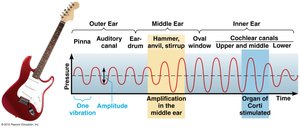
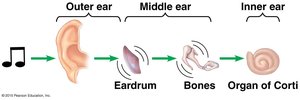
Hearing: From Sound to Action Potentials
The ear transduces sound vibrations into neural signals via the following pathway:
Outer ear: Collects sound and directs it to the eardrum.
Middle ear: Ossicles vibrate and amplify sound.
Inner ear: Cochlea converts vibrations into fluid pressure waves; organ of Corti hair cells bend, leading to transduction and action potentials.
Loudness: Encoded by action potential frequency.
Pitch: Encoded by location along the basilar membrane.
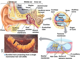
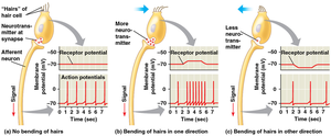
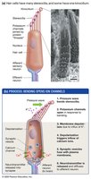
Hair Cell Transduction
Hair cells in the organ of Corti transduce mechanical stimuli into electrical signals.
Stereocilia: Actin-reinforced microvilli arranged in increasing height; tallest is the kinocilium.
Bending toward kinocilium: Opens channels, K+ influx, depolarization, Ca2+ entry, neurotransmitter release.
Key difference: Extracellular fluid is high in K+, unlike typical neurons.
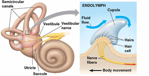
Vestibular System: Equilibrium and Balance
Vestibular System Structure and Function
The vestibular system senses head position and movement to maintain balance.
Utricle & saccule (otoliths): Detect gravity and linear acceleration.
Semicircular canals (cupula): Detect rotational movement in three planes.
Motion sickness: Caused by visual–vestibular mismatch.
Visual System: Eye Structure and Phototransduction
Vertebrate Eye Anatomy
The vertebrate eye focuses light onto photoreceptors in the retina, enabling vision.
Cornea: Transparent front surface, begins focusing.
Iris: Pigmented muscle controlling light entry.
Pupil: Opening in iris.
Lens: Focuses light onto retina.
Retina: Contains photoreceptors (rods and cones).
Blind spot: Optic disc, no photoreceptors, optic nerve exits.
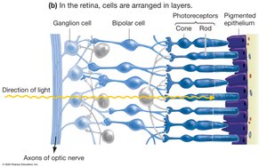
Retinal Cell Layers and Signal Pathway
Light passes through several cell layers in the retina before reaching photoreceptors.
Photoreceptors: Rods (rhodopsin) for dim light, cones (opsins) for color vision.
Bipolar cells: Relay signals from photoreceptors.
Ganglion cells: Send axons to the brain via the optic nerve.
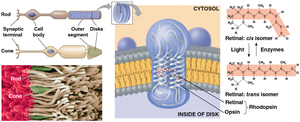
Phototransduction Mechanism
Phototransduction is the process by which light is converted into electrical signals in the retina.
Opsin (cones): Transmembrane protein packed in membranous disks.
Rhodopsin (rods): Composed of retinal and opsin.
Light absorption: Retinal changes from 11-cis to all-trans, opsin changes shape, initiating the phototransduction cascade.
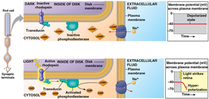
Phototransduction Cascade
Photon absorption: 11-cis retinal → all-trans retinal, rhodopsin activated.
Rhodopsin: Activates transducin (G protein), which activates phosphodiesterase (PDE).
PDE: Converts cGMP to GMP, decreasing intracellular cGMP.
Decreased cGMP: Na+/Ca2+ channels close, leading to hyperpolarization of the photoreceptor.
Processing of Visual Information
Visual Pathways and Brain Processing
Visual information is processed by the brain after being transmitted from the retina.
Optic nerves: Meet at the optic chiasm; left visual field goes to right brain, right visual field to left brain.
Lateral geniculate nucleus (LGN): Most ganglion cell axons synapse here before reaching the primary visual cortex.
~30% of cortex: Participates in visual processing.
Summary Table: Sensory Receptor Types and Functions
Receptor Type | Stimulus Detected | Example |
|---|---|---|
Mechanoreceptor | Pressure, vibration, stretch | Touch, hearing |
Photoreceptor | Light | Vision |
Chemoreceptor | Molecules | Taste, smell |
Thermoreceptor | Temperature | Heat, cold |
Nociceptor | Pain, tissue damage | Injury detection |
Electroreceptor | Electric fields | Some fish |
Magnetoreceptor | Magnetic fields | Navigation in birds |
Key Equations
Action Potential Frequency Coding
Stimulus intensity is encoded by the frequency of action potentials:
Where f is frequency, N is the number of action potentials, and T is the time interval.
Sound Wave Properties
Sound wave frequency and amplitude:
Where A is amplitude, f is frequency, t is time, and \phi is phase.
Phototransduction Cascade
cGMP conversion by phosphodiesterase:
Decreased cGMP closes Na+/Ca2+ channels.
Conclusion
Sensory and motor mechanisms are essential for animal survival, enabling detection of environmental stimuli and coordinated responses. Understanding the structure and function of sensory receptors, signal transduction, and neural coding provides insight into how animals interact with their environment. Additional info: Academic context was added to clarify mechanisms and provide self-contained explanations for exam preparation.
