 Back
BackSensory and Motor Mechanisms: Study Notes for Biology Students
Study Guide - Smart Notes
 Tailored notes based on your materials, expanded with key definitions, examples, and context.
Chapter 50: Sensory and Motor Mechanisms
Introduction to Sensory and Motor Mechanisms
Sensory and motor mechanisms are essential for animals to detect environmental stimuli and respond appropriately. These processes involve specialized cells and organs that convert various forms of energy into signals interpreted by the nervous system, resulting in coordinated motor responses. 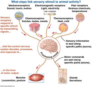
Concept 50.1: Sensory Receptors and Signal Transduction
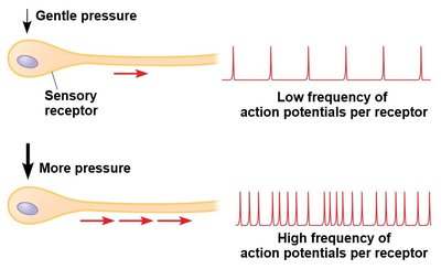
Sensory receptors are specialized cells or organs that detect stimuli and initiate the sensory pathway. They can be either neurons or non-neuronal cells and interact with both internal and external stimuli. - Sensory Reception: The process of detecting stimuli by sensory receptors. - Sensory Transduction: Conversion of stimulus energy into a change in membrane potential, known as the receptor potential. - Transmission: Sensory information is transmitted as action potentials through the nervous system. The frequency of action potentials correlates with stimulus intensity. - Perception: The brain constructs perceptions from incoming signals, distinguishing stimuli based on neural pathways. 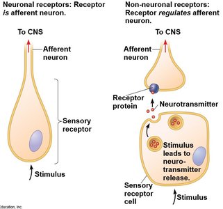
Types of Sensory Receptors
Sensory receptors are classified by the type of stimulus they detect:
Mechanoreceptors: Detect physical deformation (touch, sound, motion).
Chemoreceptors: Respond to chemical stimuli (taste, smell).
Electromagnetic Receptors: Detect light, electricity, magnetism.
Thermoreceptors: Sense heat and cold.
Pain Receptors (Nociceptors): Detect harmful conditions (excess heat, pressure, chemicals).
Concept 50.2: Mechanoreceptors in Hearing and Equilibrium
Mechanoreceptors play a crucial role in detecting moving fluid or settling particles, which is fundamental for hearing and equilibrium in animals.
Equilibrium in Invertebrates
- Most invertebrates use statocysts containing mechanoreceptors and statoliths to maintain equilibrium and sense gravity.  - Insects detect sound via body hairs and tympanic membranes.
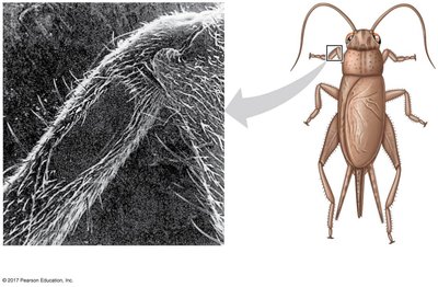
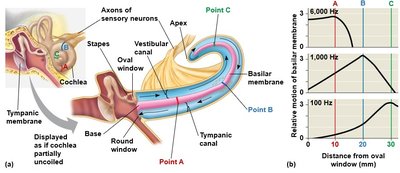
Hearing in Vertebrates
- In humans, the ear transduces pressure waves into nerve impulses using hair cells. - The ear distinguishes volume (amplitude) and pitch (frequency) via the cochlea and basilar membrane. 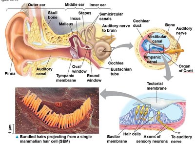
Equilibrium in Vertebrates
- The utricle and saccule contain hair cells and otoliths for sensing gravity and linear movement. - Semicircular canals detect angular movement. 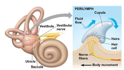
Hearing and Equilibrium in Aquatic Vertebrates
- Fishes and aquatic amphibians have a lateral line system with mechanoreceptors to detect water movement. 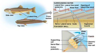
Concept 50.3: Visual Receptors and Light Detection
Animals possess diverse visual organs, but all rely on photoreceptors containing light-absorbing pigments.
Light-Detecting Organs in Invertebrates
- Planarians have simple eyespots (ocelli) to detect light direction and intensity. 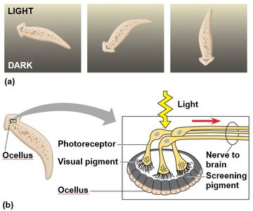
Compound Eyes
- Insects and crustaceans have compound eyes made of ommatidia, effective for detecting movement and color. 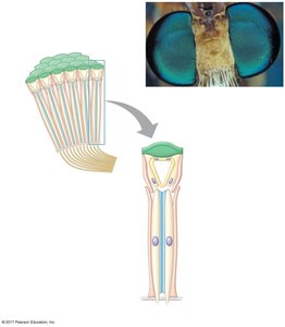
Single-Lens Eyes
- Found in vertebrates and some invertebrates, single-lens eyes function like cameras, focusing light onto the retina.
The Vertebrate Visual System
- The eye consists of layers including the choroid, retina, lens, aqueous humor, and vitreous humor. - Rods detect light intensity; cones provide color vision. 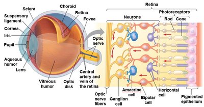
Visual Pigments and Phototransduction
- Visual pigments consist of retinal bound to opsin proteins (e.g., rhodopsin). - Light absorption changes retinal from cis to trans form, initiating signal transduction. 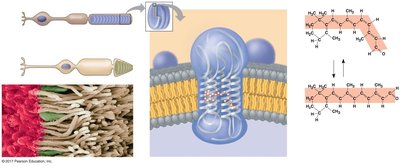
Processing Visual Information
- In darkness, rods and cones release glutamate; light causes hyperpolarization and reduces glutamate release, altering bipolar cell activity. 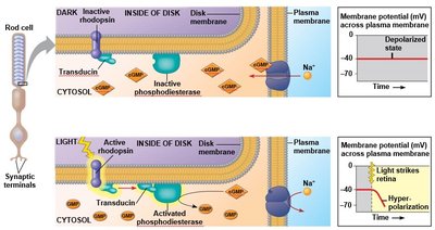
Color Vision
- Most vertebrates have good color vision; humans have three types of cones (red, green, blue) with distinct photopsins. 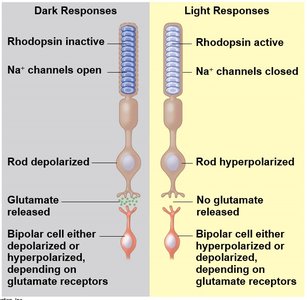
Concept 50.4: Taste and Smell
Taste (gustation) and smell (olfaction) rely on chemoreceptors to detect specific molecules.
Taste in Mammals
- Five taste perceptions: sweet, sour, salty, bitter, umami. - Taste buds contain modified epithelial cells and are located on papillae of the tongue. 
Types of Taste Receptors
- Sweet, umami, and bitter: G protein-coupled receptors (GPCRs). - Sour: TRP family receptor. - Salty: Sodium channel.
Smell in Mammals
- Olfactory receptor cells in the nasal cavity detect odorants via signal transduction, allowing mammals to distinguish thousands of odors. 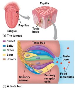
Concept 50.5: Muscle Function and Motor Response
Muscle activity is a response to nervous system input and relies on the interaction of protein filaments.
Vertebrate Skeletal Muscle Structure
- Skeletal muscle consists of bundles of fibers, each containing myofibrils. - Myofibrils are composed of repeating units called sarcomeres, bordered by Z lines. 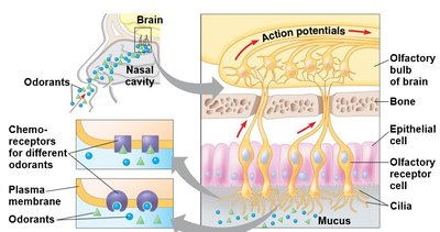
Sliding-Filament Model of Muscle Contraction
- Thin (actin) and thick (myosin) filaments slide past each other, powered by myosin heads forming cross-bridges with actin. - Muscle contraction requires cycles of binding and release, driven by ATP. 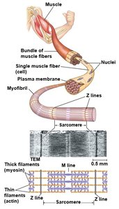
Role of Calcium and Regulatory Proteins
- Tropomyosin and troponin complex regulate actin-myosin interaction. - Calcium ions bind to troponin, exposing myosin-binding sites and enabling contraction. 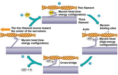
Nervous Control of Muscle Tension
- Muscle contraction is graded by varying the number of fibers contracting and the rate of stimulation. - A motor unit consists of a motor neuron and all the muscle fibers it controls.
Types of Skeletal Muscle Fibers
- Oxidative fibers: Use aerobic respiration, rich in mitochondria and myoglobin (dark meat). - Glycolytic fibers: Use glycolysis, less myoglobin, larger diameter, tire easily (white meat). - Fast-twitch fibers: Rapid, powerful contractions; can be oxidative or glycolytic. - Slow-twitch fibers: Sustained contractions; always oxidative.
Other Types of Muscle
- Cardiac muscle: Striated, found only in the heart, electrically connected by intercalated disks, can generate action potentials without neural input. - Smooth muscle: Found in walls of hollow organs, lacks striations, contractions are slow and regulated by Ca2+ via a different mechanism than skeletal muscle.
Summary Table: Types of Sensory Receptors
Receptor Type | Stimulus Detected | Example |
|---|---|---|
Mechanoreceptor | Touch, sound, motion | Hair cells in ear |
Chemoreceptor | Chemicals (taste, smell) | Taste buds, olfactory cells |
Electromagnetic receptor | Light, electricity, magnetism | Photoreceptors in eye |
Thermoreceptor | Heat, cold | Skin thermoreceptors |
Pain receptor (nociceptor) | Harmful conditions | Free nerve endings |
Key Equations
Action Potential Frequency:
Sliding Filament Model:
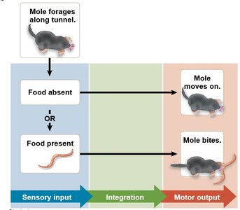
Example: Sensory Input to Motor Output
The star-nosed mole uses its specialized mechanoreceptors to detect prey in tunnels. Sensory input (touch) is integrated by the nervous system, resulting in motor output (biting or moving on). 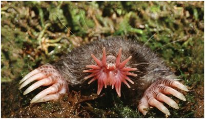
Additional info:
Some diagrams and explanations have been expanded for clarity and completeness, including the summary table and equations.
