 Back
Sensory and Motor Systems: Somatosensory Pathways and Neural Integration
Study Guide - Smart Notes
 Tailored notes based on your materials, expanded with key definitions, examples, and context.
Sensory & Motor Systems Overview
Introduction to Sensory and Motor Integration
The sensory and motor systems are essential for detecting environmental stimuli and generating appropriate responses. Sensory receptors transduce physical or chemical stimuli into electrical signals, which are processed by the nervous system to produce conscious perception and motor output.
Sensory receptors detect specific stimuli (e.g., touch, temperature, pain).
Neural pathways transmit sensory information to the central nervous system (CNS).
Motor commands are generated in response and sent to effector organs (muscles, glands).
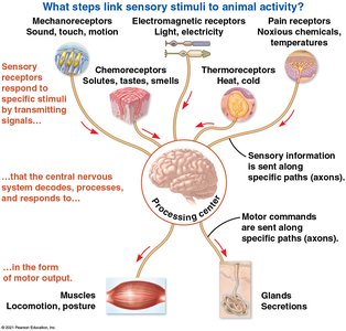
The Somatosensory System
Organization and Function
The somatosensory system is a division of the peripheral nervous system responsible for processing sensory input from the skin, muscles, and joints. It enables the detection of touch, pressure, temperature, pain, and proprioception (body position).
Stimuli are detected by sensory receptors and converted into action potentials (APs).
APs are transmitted via afferent neurons toward the CNS for processing.
Information is integrated at multiple neural levels, culminating in conscious perception.
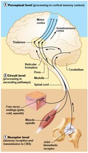
Neural Integration Levels
Receptor Level: Sensory receptors detect and transduce stimuli, generating graded potentials and, if threshold is reached, action potentials.
Circuit Level: Impulses are relayed and processed by interneurons as they ascend through the spinal cord toward the brain.
Perceptual Level: Signals reach the somatosensory cortex in the parietal lobe, where conscious awareness (perception) occurs.
The Skin and Its Receptors
Structure of the Skin
The skin, part of the integumentary system, is composed of two main layers:
Epidermis: Thin, outer layer of keratinized epithelial tissue.
Dermis: Thicker, deeper layer of connective tissue containing nerves, blood vessels, glands, and hair follicles.
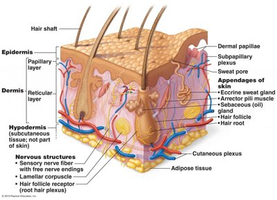
Functional Classification of Skin Receptors
Mechanoreceptors: Detect touch and pressure.
Thermoreceptors: Detect temperature changes.
Nociceptors: Detect painful stimuli (mechanical, thermal, chemical).
Proprioceptors: Detect changes in body position and movement (located in muscles and joints).
Adaptation of Sensory Receptors
Sensory receptors can be classified based on their adaptation to sustained stimuli:
Rapidly Adapting (Phasic) Receptors: Respond quickly at the onset and offset of a stimulus, useful for detecting changes (e.g., vibration, movement).
Slowly Adapting (Tonic) Receptors: Continue to fire as long as the stimulus is present, providing information about the duration and intensity of the stimulus (e.g., deep pressure).
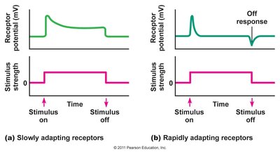
Types of Sensory Receptors in the Skin
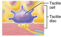
Merkel Cells (Tactile Discs)
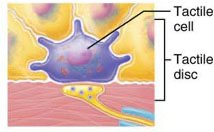
Merkel cells are specialized epithelial cells located at the base of the epidermis. They form synaptic contacts with unipolar sensory neurons, together called Merkel (tactile) discs. These receptors detect light touch and pressure and are slowly adapting.
Transduce tactile stimuli and release neurotransmitters (e.g., glutamate) to excite sensory neurons.
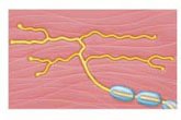
Free Nerve Endings
Free nerve endings are unencapsulated dendrites of sensory neurons found throughout the skin. They detect a variety of stimuli:
Nociceptors: Detect pain from mechanical, thermal, or chemical sources.
Thermoreceptors: Detect temperature changes; respond maximally outside the physiological zero range (where cold and warm receptor responses overlap).
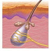
Hair Root Plexuses
These are free nerve endings that wrap around hair follicles, detecting light touch as hair moves. They are rapidly adapting receptors.
Encapsulated Receptors
Meissner (Tactile) Corpuscles: Encapsulated receptors abundant in hairless skin (fingertips, lips). Detect light touch and vibration; rapidly adapting.
Pacinian (Lamellar) Corpuscles: Large, deep receptors surrounded by layers of connective tissue. Detect deep pressure and stretch; rapidly adapting.
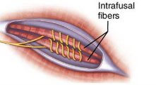
Proprioceptors
Located in muscles, tendons, and joints, these mechanoreceptors detect stretch and movement, contributing to proprioception (sense of body position).
Mechanisms of Sensory Transduction
Graded Potentials and Action Potentials
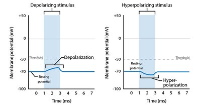
Sensory transduction begins with a change in membrane potential (graded potential) in response to a stimulus. If the graded potential reaches threshold, it triggers an action potential that travels toward the CNS.
Depolarization: Influx of cations (Na+, Ca2+) reduces the membrane potential, making the inside less negative.
Hyperpolarization: Outflow of K+ or influx of Cl- makes the inside more negative.
Transduction in Epithelial Cell Receptors (e.g., Merkel Cells)
Stimulus opens mechanically-gated cation channels, causing depolarization (receptor potential).
Depolarization opens voltage-gated Ca2+ channels, leading to neurotransmitter (glutamate) release.
Glutamate binds to ligand-gated channels on the sensory neuron, generating an excitatory postsynaptic potential (EPSP).
If the EPSP reaches threshold at the trigger zone, an action potential is generated.
Transduction in Neuronal Receptors (e.g., Pacinian Corpuscles)
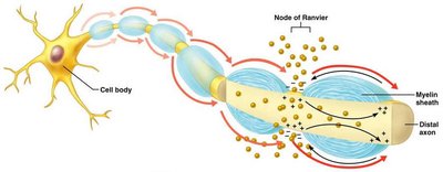
Stimulus opens mechanically-gated Na+ channels in the neuron membrane.
Na+ influx generates a generator potential (graded depolarization).
If threshold is reached at the trigger zone, an action potential is initiated and propagated toward the CNS.
Neural Pathways: From Sensation to Perception
Spinal Cord Anatomy and Sensory Pathways
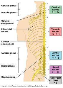
The spinal cord is protected by vertebrae, meninges, and cerebrospinal fluid. It contains gray matter (neuron cell bodies, synapses) and white matter (myelinated axons in columns called funiculi).
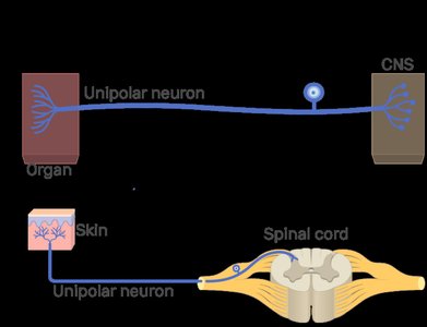
Dorsal root: Contains incoming sensory (afferent) fibers; cell bodies in dorsal root ganglia.
Ventral root: Contains outgoing motor (efferent) fibers.
Three-Neuron Ascending Pathway
Conscious sensory information typically ascends to the somatosensory cortex via a three-neuron chain:
First-order neuron: Unipolar sensory neuron; cell body in dorsal root ganglion; transduces stimulus and synapses in spinal cord or brainstem.
Second-order neuron: Multipolar interneuron; axon decussates (crosses) to contralateral side and ascends to the thalamus.
Third-order neuron: Projects from thalamus to somatosensory cortex (parietal lobe) for conscious perception.
Divergence and Signal Processing
At each synapse, signals may diverge to multiple postsynaptic neurons, allowing for redundancy and complex processing as impulses ascend to higher brain centers.
Motor Output: From Cortex to Muscle
Primary and Premotor Cortex
The frontal lobe contains the primary and premotor cortex, which plan and initiate voluntary muscle movements. Pyramidal neurons generate action potentials that descend through the spinal cord to activate motor neurons.
Axons decussate at the medulla oblongata (pyramidal decussation), controlling contralateral muscles.
α-motor neurons in the spinal cord send impulses to skeletal muscles via the ventral root.
Neuromuscular Junction
At the neuromuscular junction, the axon terminal of an α-motor neuron releases acetylcholine (ACh), which binds to ligand-gated receptors on the muscle cell membrane, triggering muscle contraction.
Summary Table: Types of Sensory Receptors in the Skin
Receptor Type | Stimulus Detected | Adaptation | Location |
|---|---|---|---|
Merkel Cells (Tactile Discs) | Light touch, pressure | Slowly adapting | Deep epidermis |
Free Nerve Endings | Pain, temperature, crude touch | Varies (mostly slowly adapting) | Epidermis, dermis |
Meissner Corpuscles | Light touch, vibration | Rapidly adapting | Dermal papillae (hairless skin) |
Pacinian Corpuscles | Deep pressure, stretch | Rapidly adapting | Deep dermis, hypodermis |
Hair Root Plexuses | Hair movement | Rapidly adapting | Hair follicles |
Proprioceptors | Muscle/joint stretch | Varies | Muscles, tendons, joints |
Key Equations
Nernst Equation (for equilibrium potential):
Ohm's Law (for membrane potential):
Additional info: This guide covers the core concepts of somatosensory and motor integration, including receptor types, neural pathways, and the mechanisms of sensory transduction and motor output. For more details on motor systems, see Chapter 49.
