 Back
BackSensory Systems: Structure, Function, and Evolution (Lecture 10)
Study Guide - Smart Notes
 Tailored notes based on your materials, expanded with key definitions, examples, and context.
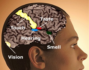
Sensory Systems Overview
Introduction to Sensory Systems
Sensory systems are specialized biological mechanisms that allow organisms to detect and interpret information from their environment. These systems rely on sensory receptors that transduce physical or chemical stimuli into neural signals, which are then processed by the nervous system to generate perception and guide behavior.
Sensory receptors are specialized cells or structures that detect specific types of stimuli (e.g., light, sound, touch, chemicals).
Transduction is the process by which sensory receptors convert external stimuli into electrical signals (receptor potentials).
These signals are transmitted to the central nervous system (CNS) for integration and interpretation.
Types of Sensory Receptors
Classification of Sensory Receptors
Sensory receptors are classified based on the type of stimulus they detect:
Mechanoreceptors: Detect mechanical forces such as touch, pressure, vibration, and stretch.
Photoreceptors: Detect light (vision).
Chemoreceptors: Detect chemical stimuli (taste and smell).
Thermoreceptors: Detect temperature changes (heat and cold).
Nociceptors: Detect pain (damaging stimuli).
Electromagnetic receptors: Detect electromagnetic fields (e.g., magnetic field detection in some animals).
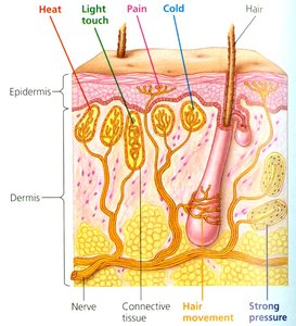
Mechanoreception: Touch and Skin Sensations
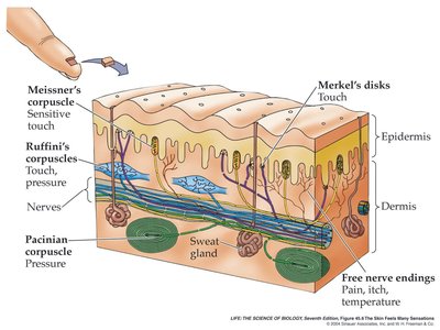
Structure and Function of Skin Receptors
The skin contains various mechanoreceptors that detect different types of mechanical stimuli. These receptors are distributed in both the epidermis and dermis and are responsible for sensations such as touch, pressure, vibration, and temperature.
Meissner's corpuscles: Sensitive to light touch, adapt rapidly.
Merkel's disks: Provide continuous information about pressure, adapt slowly.
Ruffini corpuscles: Detect skin stretch and low-frequency vibration, adapt slowly.
Pacinian corpuscles: Detect deep pressure and high-frequency vibration, adapt quickly.
Free nerve endings: Detect pain, itch, and temperature changes.
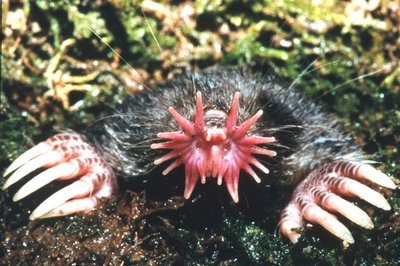
Specialized Touch Adaptations
Certain animals have evolved specialized structures with high densities of mechanoreceptors for enhanced tactile sensation. For example, the star-nosed mole possesses appendages with an extraordinary concentration of touch receptors, allowing it to detect prey efficiently in darkness.
Mechanoreception: Hearing
Hair Cells in the Cochlea
Hair cells are specialized mechanoreceptors located in the cochlea of the inner ear. They detect sound vibrations and convert them into electrical signals that are transmitted to the brain via the auditory nerve.
Sound waves cause movement of the basilar membrane, bending the hair cells.
Bending opens ion channels, generating receptor potentials and action potentials in auditory neurons.
Photoreception: Vision
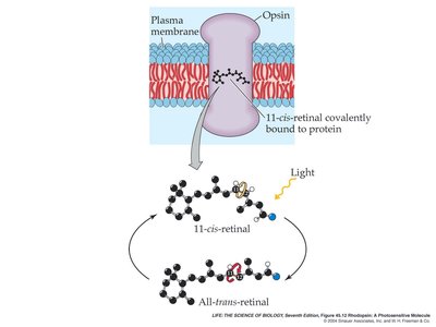
Photoreceptor Molecules and Signal Transduction
Photoreceptors are specialized cells that detect light. The primary photoreceptor molecule in animals is rhodopsin, which consists of a light-absorbing pigment (retinal) bound to a protein (opsin). When light is absorbed, retinal changes from the 11-cis to the all-trans form, triggering a conformational change in opsin and initiating a signal transduction cascade.
This cascade ultimately alters the membrane potential of the photoreceptor cell, leading to changes in neurotransmitter release.
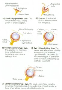
Evolution of Photoreceptors and Eyes
The evolution of eyes demonstrates a progression from simple light-sensitive patches to complex camera-type eyes. This evolutionary sequence is evident in mollusks, which display a range of eye complexities, supporting the theory of natural selection.
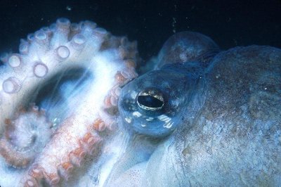
Photoreceptor Diversity and Adaptation
Some animals, such as the Eastern American mole, have vestigial eyes due to relaxed selective pressure for vision in their environment, illustrating the concept of "de-evolution." In contrast, cephalopods and vertebrates have highly evolved eyes, though their anatomical designs differ.

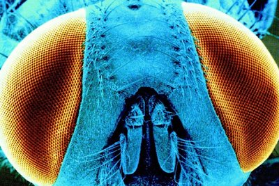
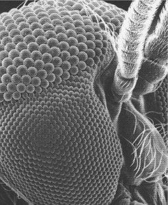
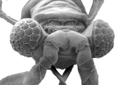
Compound Eyes in Arthropods
Arthropods, such as insects, possess compound eyes composed of numerous ommatidia, each functioning as an individual photoreceptive unit. This structure provides a wide field of view and sensitivity to motion, though the resulting image is less sharp than that produced by vertebrate eyes.
Each ommatidium contains a lens and photoreceptor cells (retinula cells) with rhodopsin.
The number of ommatidia varies among species, influencing visual acuity.
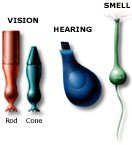
Vertebrate Eye Structure and Function
The vertebrate eye contains two main types of photoreceptors: rods and cones. Rods are highly sensitive to low light and provide black-and-white vision, while cones are responsible for color vision and visual acuity. The fovea, a region of the retina, contains a high density of cones and is the site of sharpest vision.
Light absorption by rhodopsin in rods triggers a G protein (transducin) cascade, leading to closure of Na+ channels and hyperpolarization of the cell.
In the dark, photoreceptors are depolarized and release glutamate; in the light, they hyperpolarize and reduce glutamate release.
Bipolar cells and ganglion cells process and transmit visual information to the brain via the optic nerve.
Color Vision
Humans have three types of cone cells, each sensitive to different wavelengths (blue, green, red). The combination of signals from these cones allows for the perception of a wide range of colors. Other animals may have different numbers of cone types, resulting in dichromatic, trichromatic, or tetrachromatic vision.
Limits and Extensions of Sensory Perception
The World Beyond Human Perception
Many animals possess sensory capabilities beyond those of humans, such as detection of ultraviolet or infrared light, electric fields, echolocation, and magnetic fields. Sensory systems are adapted to the ecological needs of each species, emphasizing survival-relevant information rather than an objective representation of reality.
Examples: Bees see ultraviolet patterns on flowers; snakes detect infrared radiation; bats use echolocation.
Philosophical Implications
Perception and Reality
Sensory systems construct a neural representation of the environment, which may differ from objective reality. Philosophers such as Plato, Descartes, Berkeley, and Kant have explored the implications of perception and the limits of human knowledge.
Practice Questions
Multiple Choice Example
Consider a sensory receptor such as a stretch receptor in a vertebrate muscle: as compared with a weak stimulus that barely reaches above threshold, a very strong stimulus evokes in the receptor cell’s axon:
A) A higher frequency of action potentials
B) A lower frequency of action potentials
C) A more depolarized action potential as it progresses along the axon
D) A more hyperpolarized action potential as it progresses along the axon
E) A change from passive flow of current to salutatory conduction
F) More than one of the above (A-E).
G) None of the above (A-E).
Short Answer Example
One of the potential side effects of Viagra is “a sudden loss of vision.” Given your understanding of the enzyme which is the target of Viagra in the case of penile erection, explain why it is not surprising that Viagra might also have an effect on vision. Your answer should include:
A. a complete description of the signal transduction pathway in the photoreceptors of the visual system.
B. an explanation of where and how in this pathway Viagra might be causing problems.
Additional info: This guide integrates and expands upon the provided lecture notes, including definitions, examples, and academic context for completeness.
