 Back
Structure and Function of Large Biological Molecules: Proteins & Nucleic Acids
Study Guide - Smart Notes
 Tailored notes based on your materials, expanded with key definitions, examples, and context.
Proteins: Structure and Function
Overview of Proteins
Proteins are essential macromolecules that perform a vast array of functions within cells. They account for more than 50% of the dry mass of most cells and are encoded by thousands of genes, with post-translational modifications and alternative splicing increasing their diversity into the millions.
Proteins are polymers made from 20 different amino acids.
Each protein has a unique three-dimensional shape, which determines its function.
Proteins are involved in nearly all cellular processes.
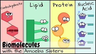
Protein Functions
Proteins serve diverse functions in biological systems. The eight major categories of protein functions include:
Enzymatic proteins: Catalyze biochemical reactions (e.g., DNA polymerase).
Defensive proteins: Protect against disease (e.g., antibodies).
Storage proteins: Store amino acids (e.g., casein in milk).
Transport proteins: Move substances (e.g., hemoglobin transports oxygen).
Hormonal proteins: Coordinate organismal activities (e.g., insulin regulates blood sugar).
Receptor proteins: Respond to chemical signals (e.g., nerve cell receptors).
Contractile and motor proteins: Enable movement (e.g., actin and myosin in muscles).
Structural proteins: Provide support (e.g., collagen in connective tissue).
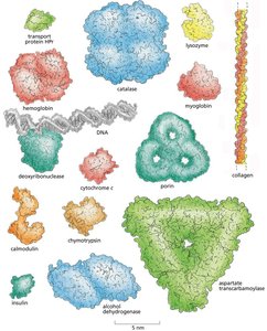
Enzymes
Enzymes are proteins that act as catalysts, speeding up chemical reactions without being consumed.
Catalysts lower the activation energy required for reactions.
Enzymes are reusable and highly specific for their substrates.
Proteins as Polypeptides
Proteins are polymers called polypeptides, made by linking amino acids via peptide bonds.
Amino acids are the monomers of proteins.
All organisms use the same set of 20 amino acids.
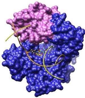
A functional protein may consist of one or more polypeptides folded into a specific shape.
Amino Acids: Structure and Properties
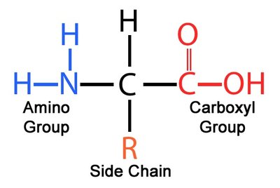
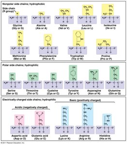
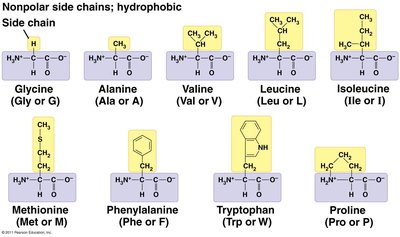
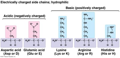
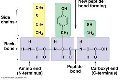
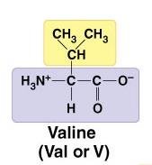
Amino acids have a central carbon atom bonded to an amino group, a carboxyl group, a hydrogen atom, and a variable side chain (R group). The side chain determines the properties of each amino acid.
There are 20 different amino acids, each with a unique R group.
Amino acids are classified as hydrophobic (nonpolar), hydrophilic (polar), or charged (acidic/basic).
Formation of Peptide Bonds
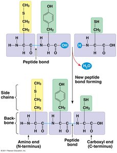
Amino acids are joined by dehydration reactions, forming peptide bonds.
The peptide bond links the carboxyl group of one amino acid to the amino group of another.
Breaking a peptide bond is a hydrolysis reaction.
Protein Orientation: N-terminus and C-terminus
The backbone of a protein is a chain of peptide bonds.
The first amino acid has a free amino group (N-terminus).
The last amino acid has a free carboxyl group (C-terminus).
New amino acids are always added to the C-terminus.
Levels of Protein Structure
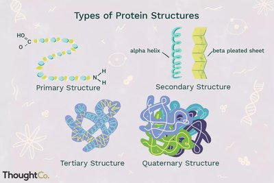
Proteins fold into four levels of structure, each critical for function.
Primary structure: Sequence of amino acids.
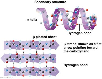
Secondary structure: Local folding into α-helices and β-sheets, stabilized by hydrogen bonds.
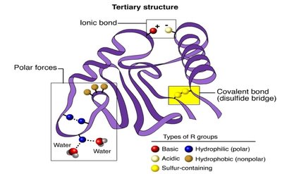
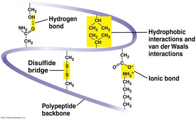
Tertiary structure: Overall 3D shape, determined by interactions among side chains (R groups).
Quaternary structure: Association of multiple polypeptide chains.
Protein Structure Summary Table
Level | Description | Stabilizing Bonds |
|---|---|---|
Primary | Amino acid sequence | Peptide bonds |
Secondary | α-helix, β-sheet | Hydrogen bonds (backbone) |
Tertiary | 3D folding | Hydrogen bonds, ionic bonds, hydrophobic interactions, disulfide bridges (side chains) |
Quaternary | Multiple polypeptides | Same as tertiary, between chains |
Protein Folding and Chaperonins
Protein folding is often spontaneous but can be assisted by chaperone proteins called chaperonins.
Chaperonins provide a protective environment for proper folding.
Correct folding is essential for protein function.
Denaturation of Proteins
Most bonds stabilizing protein structure (secondary, tertiary, quaternary) are weak and can be disrupted by changes in pH, temperature, or salt concentration.
Denatured proteins lose their functional shape.
Proteins may refold if the environment is restored.
Sickle-Cell Disease: A Case Study
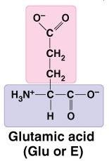
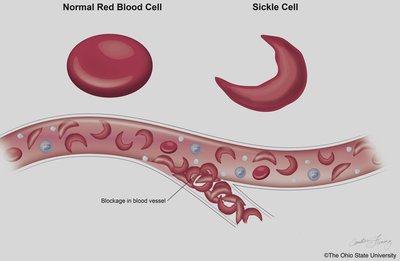
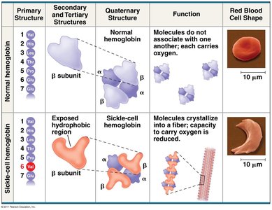
Sickle-cell disease is caused by a single amino acid substitution in the primary structure of hemoglobin (glutamic acid replaced by valine).
This change alters the protein's shape and the red blood cell's shape.
Demonstrates how primary structure affects higher-order structure and function.

Nucleic Acids: Structure and Function
Overview of Nucleic Acids
Nucleic acids are the fourth major class of macromolecules and serve as the genetic material of cells.
Monomer: Nucleotide
Polymers: DNA (deoxyribonucleic acid) and RNA (ribonucleic acid)
DNA stores hereditary information; genes are specific nucleotide sequences that code for proteins.
RNA assists in protein production.
Flow of Genetic Information
The flow of genetic information in cells follows the central dogma: DNA → RNA → Protein.
DNA is transcribed into RNA.
RNA is translated into protein.
Summary Table: Macromolecules
Macromolecule | Monomer | Polymer | Main Function |
|---|---|---|---|
Protein | Amino acid | Polypeptide | Cellular functions, structure, enzymes |
Nucleic Acid | Nucleotide | DNA, RNA | Genetic information, protein synthesis |
Additional info:
Alternative splicing and post-translational modifications greatly increase protein diversity.
Module 4 will cover DNA, RNA, and their roles in protein production in detail.
