 Back
Structure and Function of Large Biological Molecules: Study Notes
Study Guide - Smart Notes
 Tailored notes based on your materials, expanded with key definitions, examples, and context.
The Structure and Function of Large Biological Molecules
Overview: The Molecules of Life
All living organisms are composed of four major classes of large biological molecules: carbohydrates, lipids, proteins, and nucleic acids. These molecules are essential for cellular structure and function. Macromolecules are large molecules made up of thousands of covalently connected atoms, and their structure is closely related to their function.
Macromolecules: Polymers and Monomers
Polymers and Monomers
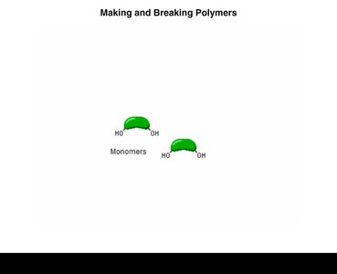
A polymer is a long molecule consisting of many similar building blocks called monomers. Three of the four classes of life’s organic molecules—carbohydrates, proteins, and nucleic acids—are polymers.
Synthesis and Breakdown of Polymers
Polymers are synthesized and broken down by specific chemical reactions:
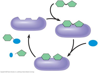
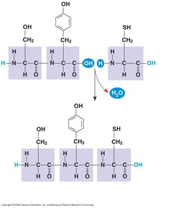
Dehydration Reaction (Condensation Reaction): Joins two monomers by removing a water molecule, forming a new bond. Enzymes catalyze this process.
Hydrolysis: Breaks a polymer into monomers by adding a water molecule, essentially the reverse of dehydration.
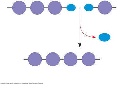
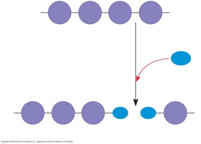
Example: Making and breaking polymers involves monomers joining or separating via these reactions.
Carbohydrates: Fuel and Building Material
Monosaccharides, Disaccharides, and Polysaccharides
Carbohydrates include sugars and their polymers. The simplest carbohydrates are monosaccharides (single sugars), which serve as major fuel for cells and as raw material for building molecules. Polysaccharides are carbohydrate macromolecules composed of many sugar building blocks.
Monosaccharides have molecular formulas that are usually multiples of CH2O.
Glucose (C6H12O6) is the most common monosaccharide.
Monosaccharides are classified by the location of the carbonyl group (aldose or ketose) and the number of carbons in the skeleton.
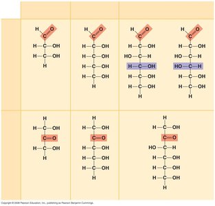
In aqueous solutions, many sugars form rings rather than linear skeletons.
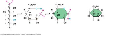
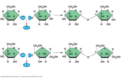
Formation of Disaccharides and Polysaccharides
A disaccharide is formed when a dehydration reaction joins two monosaccharides, creating a glycosidic linkage. Polysaccharides have storage and structural roles, determined by their sugar monomers and the positions of glycosidic linkages.
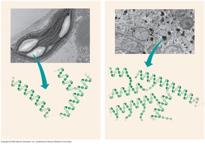
Storage Polysaccharides
Starch: Storage polysaccharide in plants, consisting entirely of glucose monomers. Stored as granules in chloroplasts and other plastids.
Glycogen: Storage polysaccharide in animals, mainly in liver and muscle cells.
Structural Polysaccharides
Cellulose: Major component of plant cell walls. Like starch, it is a polymer of glucose, but with different glycosidic linkages (alpha vs beta forms).
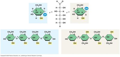
Cellulose molecules are grouped into microfibrils, forming strong building materials for plants.
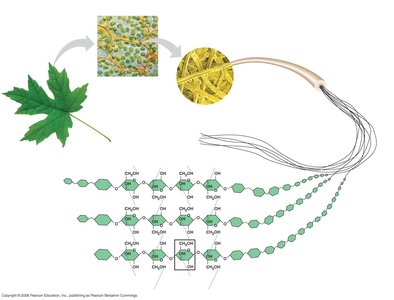
Enzymes that digest starch (alpha linkages) cannot hydrolyze beta linkages in cellulose.
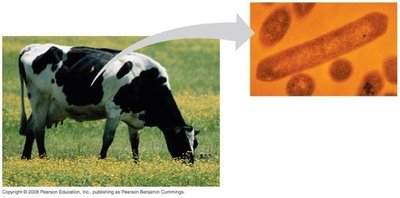
Cellulose in human food passes as insoluble fiber; some microbes can digest cellulose.
Herbivores have symbiotic relationships with microbes that digest cellulose.
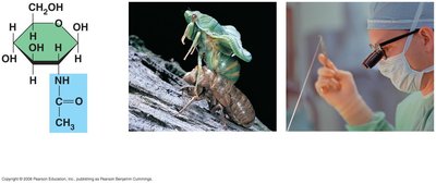
Chitin: Structural polysaccharide found in arthropod exoskeletons and fungal cell walls. Used in surgical thread for its strength and flexibility.
Lipids: Hydrophobic Molecules
Fats, Phospholipids, and Steroids
Lipids are a diverse group of hydrophobic molecules that do not form polymers. Their main types are fats, phospholipids, and steroids. Fats are constructed from glycerol and fatty acids, and their major function is energy storage.
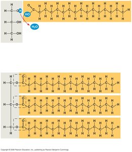
Fats are made from glycerol (a three-carbon alcohol) and fatty acids (carboxyl group attached to a long carbon skeleton).
Three fatty acids join to glycerol by ester linkage, forming a triacylglycerol (triglyceride).
Saturated fatty acids have no double bonds; unsaturated fatty acids have one or more double bonds.
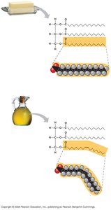
Saturated fats are solid at room temperature (animal fats); unsaturated fats are liquid (plant and fish fats).
Hydrogenation converts unsaturated fats to saturated fats and can create trans fats, which may contribute to cardiovascular disease.
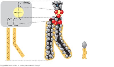
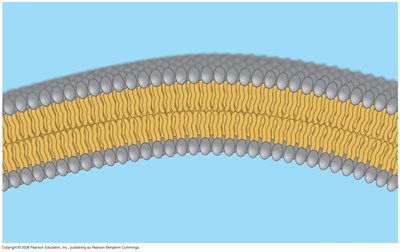
Phospholipids: Two fatty acids and a phosphate group attached to glycerol. Hydrophobic tails and hydrophilic head allow phospholipids to form bilayers in cell membranes.
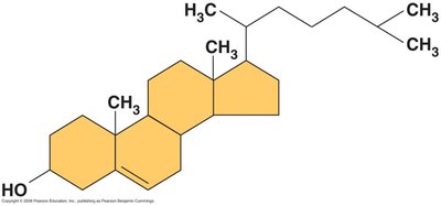
Steroids: Lipids with a carbon skeleton of four fused rings. Cholesterol is an essential steroid in animal cell membranes.
Proteins: Structure and Function
Protein Functions
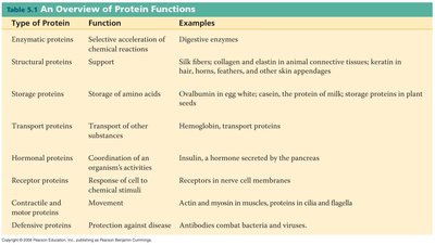
Proteins account for more than 50% of the dry mass of most cells and serve diverse functions, including structural support, storage, transport, cellular communication, movement, and defense.
Type of Protein | Function | Examples |
|---|---|---|
Enzymatic proteins | Selective acceleration of chemical reactions | Digestive enzymes |
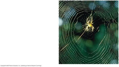
Structural proteins | Support | Silk, collagen, elastin, keratin |
Storage proteins | Storage of amino acids | Ovalbumin, casein |
Transport proteins | Transport of substances | Hemoglobin |
Hormonal proteins | Coordination of activities | Insulin |
Receptor proteins | Response to stimuli | Receptors in nerve cells |
Contractile and motor proteins | Movement | Actin, myosin |
Defensive proteins | Protection against disease | Antibodies |
Polypeptides and Amino Acids
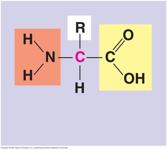
A protein consists of one or more polypeptides, which are polymers built from 20 amino acids. Amino acids have carboxyl and amino groups and differ by their side chains (R groups).
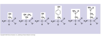
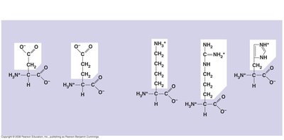
Amino acids are linked by peptide bonds, forming polypeptides with a unique linear sequence.
Protein Structure
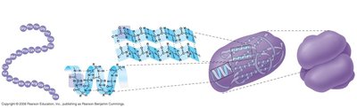
Protein structure is hierarchical:
Primary structure: Unique sequence of amino acids.
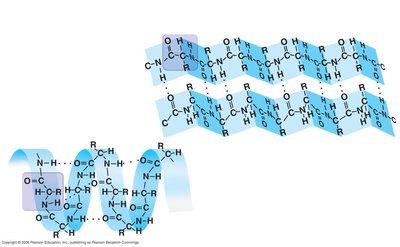
Secondary structure: Coils and folds (alpha helix, beta pleated sheet) due to hydrogen bonding.
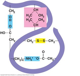
Tertiary structure: Interactions among R groups (hydrogen bonds, ionic bonds, hydrophobic interactions, disulfide bridges).
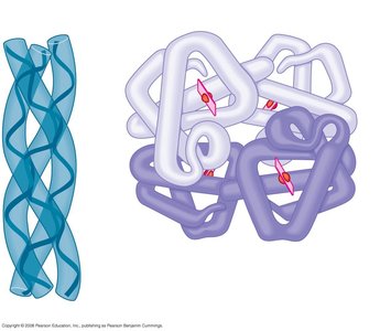
Quaternary structure: Multiple polypeptide chains forming one macromolecule (e.g., collagen, hemoglobin).
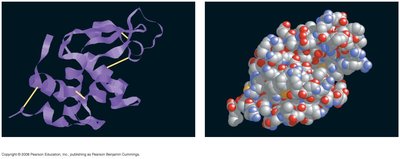
Sickle-Cell Disease: Change in Primary Structure
A single amino acid substitution in hemoglobin can cause sickle-cell disease, affecting protein function and cell shape.
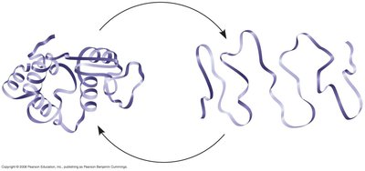
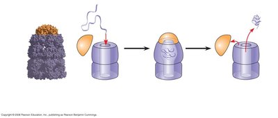
Denaturation: Loss of a protein’s native structure due to changes in pH, salt concentration, or temperature.
Chaperonins: Protein molecules that assist the proper folding of other proteins.
Nucleic Acids: Hereditary Information
DNA and RNA
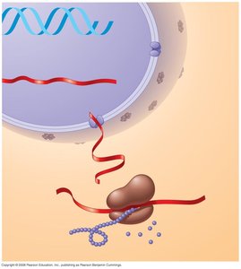
The amino acid sequence of a polypeptide is programmed by genes made of DNA. DNA directs synthesis of messenger RNA (mRNA), which controls protein synthesis in ribosomes.
Structure of Nucleic Acids
Nucleic acids are polymers called polynucleotides, made of monomers called nucleotides. Each nucleotide consists of a nitrogenous base, a pentose sugar, and a phosphate group. Nucleoside = nitrogenous base + sugar.
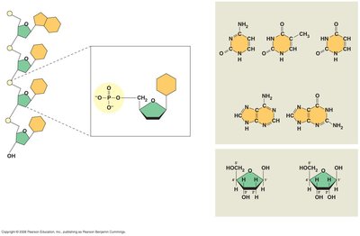
Pyrimidines: Cytosine, thymine (DNA), uracil (RNA) – single ring.
Purines: Adenine, guanine – double ring.
DNA sugar: deoxyribose; RNA sugar: ribose.
Nucleotide Polymers and DNA Double Helix
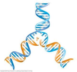
Nucleotide polymers are linked by covalent bonds between the 3' OH of one nucleotide and the 5' phosphate of the next, creating a sugar-phosphate backbone. DNA molecules have two polynucleotides forming a double helix, with antiparallel strands and specific base pairing (A-T, G-C).
Emergent Properties in the Chemistry of Life
Higher levels of organization result in the emergence of new properties. Organization is key to the chemistry of life.
Summary Table: Key Distinctions
Pair | Distinction |
|---|---|
Monosaccharide, Disaccharide, Polysaccharide | Single sugar, two sugars, many sugars |
Saturated vs Unsaturated Fat | No double bonds (solid) vs one or more double bonds (liquid) |
Cis vs Trans Fat | Cis: R groups same side; Trans: R groups opposite sides |
Pyrimidine vs Purine | Single ring vs double ring |
Nucleotide vs Nucleoside | Nucleotide: base + sugar + phosphate; Nucleoside: base + sugar |
Ribose vs Deoxyribose | Ribose: RNA sugar; Deoxyribose: DNA sugar |
5' end vs 3' end | 5' end: phosphate; 3' end: hydroxyl |
