Back
CH 17 p. 2: Structure and Function of tRNA and Ribosomes: Mechanisms of Translation
Study Guide - Smart Notes
 Tailored notes based on your materials, expanded with key definitions, examples, and context.
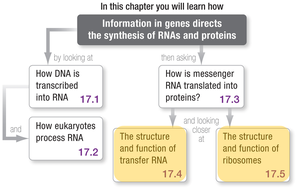
Structure and Function of Transfer RNA (tRNA)
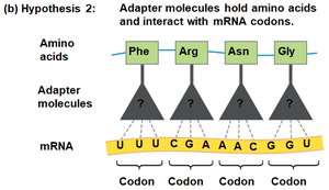
Discovery of tRNA – The Adapter Molecule
The discovery of transfer RNA (tRNA) was pivotal in understanding how genetic information is translated into proteins. tRNA was identified accidentally during experiments designed to reconstitute protein synthesis in vitro. Researchers found that, in addition to ribosomes, mRNA, amino acids, ATP, and GTP, an unknown RNA fraction was necessary for protein synthesis. This RNA was later identified as tRNA, which acts as an adapter molecule, linking amino acids to their corresponding codons on mRNA.
Function of tRNA – Experimental Evidence
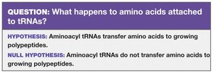
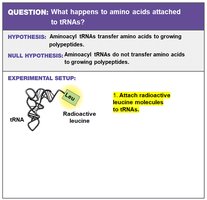
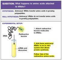
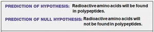
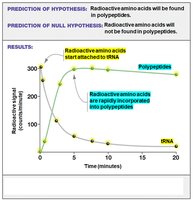
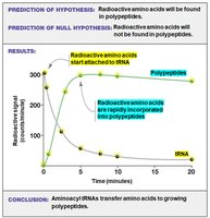
The function of tRNA was also discovered by accident. In experiments using radiolabeled leucine, researchers observed that radioactive leucine became attached to tRNAs. Further experiments showed that these aminoacyl-tRNAs could transfer their amino acids to growing polypeptides, confirming tRNA's role in translation.
Structure of tRNA
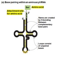
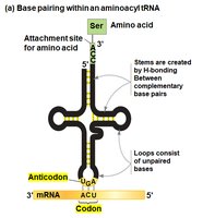
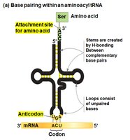
tRNAs have a characteristic secondary structure known as the cloverleaf model, formed by hydrogen bonding between complementary bases. The 3' end of the tRNA is the amino acid attachment site, while the loop opposite this site contains the anticodon, which base-pairs with the codon on the mRNA. The precise distance between the amino acid attachment site and the anticodon is critical for accurate translation.

Attachment of Amino Acids to tRNAs
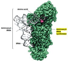
The enzyme aminoacyl-tRNA synthetase catalyzes the attachment of amino acids to their corresponding tRNAs. There is a specific synthetase for each amino acid, and the reaction requires energy from ATP. This process ensures that each tRNA carries the correct amino acid corresponding to its anticodon.
Reaction:
Structure and Function of Ribosomes
Ribosome Structure: Form is Related to Function
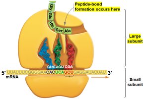
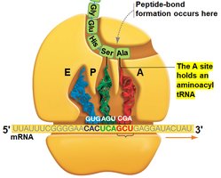
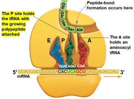
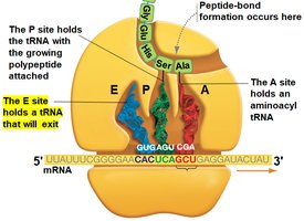
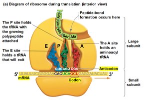
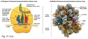
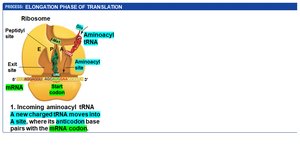
Ribosomes are large complexes of proteins and ribosomal RNAs (rRNAs), organized into two subunits: the small subunit, which binds and moves along the mRNA, and the large subunit, which catalyzes peptide bond formation. Ribosomes contain three binding sites for tRNAs:
A site (Aminoacyl site): Binds incoming aminoacyl-tRNA.
P site (Peptidyl site): Holds the tRNA with the growing polypeptide chain.
E site (Exit site): Binds uncharged tRNA ready to exit the ribosome.

Details of Bacterial Translation
Initiation Phase
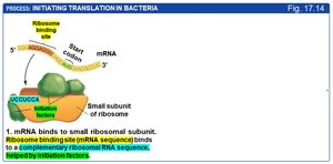
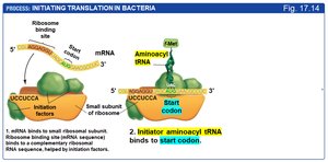
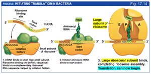
Translation initiation in bacteria involves several steps:
Initiation factors bind to the small ribosomal subunit.
rRNA in the small subunit binds to the ribosome binding sequence on the mRNA (upstream of the start codon).
The initiator tRNA binds to the start codon (AUG).
The large ribosomal subunit binds, completing the initiation complex. The initiator tRNA is positioned in the P site.
Elongation Phase
During elongation, amino acids are added sequentially to the growing polypeptide chain:
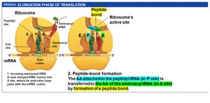
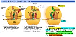
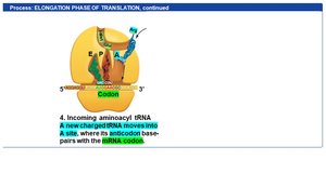
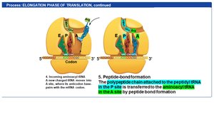
Aminoacyl-tRNAs enter the A site.
If the anticodon matches the mRNA codon, the tRNA binds.
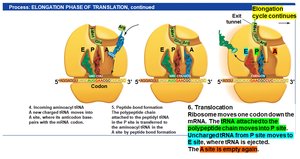
A peptide bond forms between the amino acid in the A site and the polypeptide in the P site, transferring the chain to the A site tRNA.
Translocation occurs: the ribosome moves one codon along the mRNA, shifting tRNAs to the next site (A to P, P to E, E exits).
Termination Phase
Translation ends when a stop codon is reached:
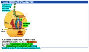
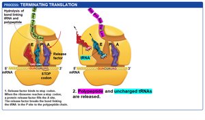
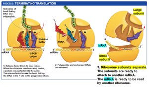
A release factor protein binds to the stop codon at the A site.
This catalyzes the hydrolysis of the bond linking the polypeptide to the tRNA in the P site.
The polypeptide and uncharged tRNAs are released, and the ribosomal subunits dissociate from the mRNA.
Post-Translational Modifications
After translation, proteins often undergo post-translational modifications to become fully functional:
Folding: Secondary structures form spontaneously; tertiary and quaternary structures may require molecular chaperones.
Chemical modifications: Addition of carbohydrates, lipids, or phosphate groups (e.g., by kinases) can occur in the rough endoplasmic reticulum or Golgi apparatus.
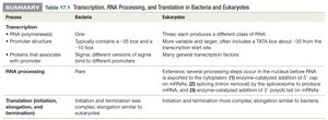
Summary Table: Transcription, RNA Processing, and Translation in Bacteria and Eukaryotes
The following table compares key aspects of gene expression in bacteria and eukaryotes:
Process | Bacteria | Eukaryotes |
|---|---|---|
RNA polymerases & promoter structure | One; typically contains a -10 box and -35 box; different versions of sigma bind to different promoters | Three; each produces a different class of RNA; more variable and larger; often includes a TATA box about -30 from the start site; many general transcription factors |
RNA processing | Rare | Extensive; several processing steps occur in the nucleus before RNA is exported to the cytoplasm (5' cap, splicing, 3' poly(A) tail) |
Translation (initiation, elongation, termination) | Initiation and termination less complex; elongation similar to eukaryotes | Initiation and termination more complex; elongation similar to bacteria |