 Back
BackStudy Guidance for Animal Form, Function, and Regulation
Study Guide - Smart Notes
 Tailored notes based on your materials, expanded with key definitions, examples, and context.
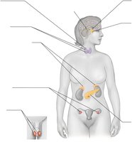
Q14. Label each of the indicated endocrine glands, name the hormone(s) it produces, and the effect of each hormone.
Background
Topic: Endocrine System Anatomy and Physiology
This question tests your understanding of the locations, hormones, and functions of the major endocrine glands in the human body.
Key Terms and Concepts:
Endocrine gland: A gland that releases hormones directly into the bloodstream.
Hormone: A chemical messenger that regulates physiological processes.
Target tissue: The specific cells or organs affected by a hormone.
Step-by-Step Guidance
Examine the diagram and identify the labeled locations of each endocrine gland (e.g., pineal, pituitary, thyroid, parathyroid, adrenal, pancreas, ovaries, testicles).
For each gland, recall or look up the primary hormone(s) it produces (e.g., insulin from pancreas, thyroxine from thyroid, cortisol from adrenal glands).
Describe the main effect of each hormone on the body (e.g., insulin lowers blood glucose, thyroxine regulates metabolism).
Consider how the hormones interact with their target tissues to produce physiological responses.
Try solving on your own before revealing the answer!
Final Answer:
The diagram shows the locations of the major endocrine glands: pineal body, pituitary gland, hypothalamus, thyroid, parathyroid, adrenal glands, pancreas, ovaries, and testicles. Each gland produces specific hormones (e.g., pituitary produces growth hormone, thyroid produces thyroxine, adrenal produces cortisol and adrenaline, pancreas produces insulin and glucagon, ovaries produce estrogen and progesterone, testicles produce testosterone). The effects of these hormones include regulation of metabolism, growth, stress response, blood sugar, and reproductive functions.
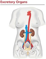
Q60. Using Figure 32.21, label all the excretory organs and major blood vessels of the figure that follows.
Background
Topic: Human Excretory System Anatomy
This question is about identifying the main organs and blood vessels involved in excretion in humans.
Key Terms:
Kidney: Filters blood and produces urine.
Ureter: Carries urine from kidney to bladder.
Bladder: Stores urine.
Urethra: Releases urine from the body.
Renal artery/vein: Blood vessels supplying and draining the kidneys.
Step-by-Step Guidance
Look at the diagram and identify the kidneys, ureters, bladder, and urethra.
Locate the major blood vessels: descending aorta, renal arteries, and renal veins.
Label each structure and note its function in the excretory process.
Consider how blood flows through the kidneys and how urine is transported and stored.
Try solving on your own before revealing the answer!
Final Answer:
The diagram shows the kidneys (bean-shaped organs), ureters (tubes connecting kidneys to bladder), bladder (urine storage), and urethra (urine exit). The descending aorta supplies blood to the kidneys via renal arteries, and renal veins drain blood from the kidneys.
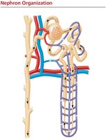
Q64. Label the following structures: glomerulus, Bowman's capsule, proximal tubule, loop of Henle, collecting duct, and renal pelvis. Next, label the following blood vessels: afferent arterioles, efferent arterioles, peritubular capillaries, and vasa recta.
Background
Topic: Nephron Structure and Blood Flow
This question tests your knowledge of nephron anatomy and the associated blood vessels in the kidney.
Key Terms:
Nephron: Functional unit of the kidney.
Glomerulus: Capillary tuft for filtration.
Bowman's capsule: Surrounds glomerulus, collects filtrate.
Proximal tubule, loop of Henle, collecting duct, renal pelvis: Segments of nephron for processing filtrate.
Afferent/efferent arterioles, peritubular capillaries, vasa recta: Blood vessels involved in filtration and reabsorption.
Step-by-Step Guidance
Examine the nephron diagram and identify each labeled structure (glomerulus, Bowman's capsule, etc.).
Trace the path of filtrate through the nephron: from glomerulus to Bowman's capsule, then through proximal tubule, loop of Henle, distal tubule, collecting duct, and finally renal pelvis.
Identify the blood vessels: afferent arteriole brings blood to glomerulus, efferent arteriole carries blood away, peritubular capillaries surround tubules, vasa recta runs alongside loop of Henle.
Label each structure and vessel, noting their role in filtration, reabsorption, and secretion.
Try solving on your own before revealing the answer!
Final Answer:
The diagram shows the nephron with glomerulus, Bowman's capsule, proximal tubule, loop of Henle, collecting duct, and renal pelvis. Blood vessels include afferent and efferent arterioles, peritubular capillaries, and vasa recta, each playing a role in filtration and reabsorption.
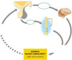
Q74. Use the figure below to describe the role of antidiuretic hormone (ADH) in maintaining blood osmolarity. Add and label the location of aquaporins.
Background
Topic: Hormonal Regulation of Water Balance
This question focuses on how ADH regulates blood osmolarity and the role of aquaporins in water reabsorption.
Key Terms:
ADH (antidiuretic hormone): Hormone that increases water reabsorption in kidneys.
Blood osmolarity: Concentration of solutes in blood.
Aquaporins: Water channel proteins in kidney tubules.
Step-by-Step Guidance
Study the diagram showing the feedback loop for blood osmolarity regulation.
Identify the role of osmoreceptors in the hypothalamus and how they stimulate ADH release from the pituitary gland.
Describe how ADH increases the permeability of the distal tubule and collecting duct by inserting aquaporins, allowing more water to be reabsorbed.
Explain how this process helps maintain normal blood osmolarity and prevents dehydration.
Try solving on your own before revealing the answer!
Final Answer:
ADH is released in response to high blood osmolarity, increasing aquaporin channels in kidney tubules, which enhances water reabsorption and restores osmolarity to normal levels.
