 Back
The Cardiovascular System: Structure and Function
Study Guide - Smart Notes
 Tailored notes based on your materials, expanded with key definitions, examples, and context.
Cardiovascular System Overview
Components and Functions
The cardiovascular system is essential for the transport of substances throughout the body, maintenance of homeostasis, and protection against disease. It consists of the heart, blood vessels (arteries, veins, and capillaries), and blood.
Transport: Delivers oxygen, carbon dioxide, nutrients, hormones, and metabolic wastes.
Homeostasis: Regulates body temperature, pH, and fluid balance.
Protection: Provides immune defense and clotting mechanisms.
Support: Maintains tissue supply and removes toxins.
The Heart
Gross Anatomy of the Heart
The heart is a muscular organ responsible for pumping blood through the systemic and pulmonary circuits. It is divided into four chambers: right atrium, right ventricle, left atrium, and left ventricle. Major vessels include the aorta, pulmonary arteries, pulmonary veins, and vena cavae.
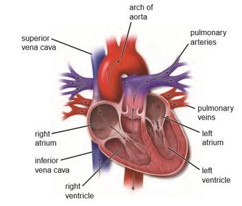
Layers of the Heart Wall
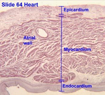
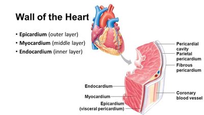
The heart wall is composed of three distinct layers, each with specialized structure and function:
Epicardium: Outer layer, also known as the visceral pericardium.
Myocardium: Middle, muscular layer responsible for contraction.
Endocardium: Inner lining in contact with blood.
Epicardium
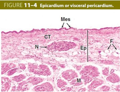
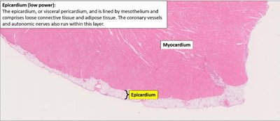
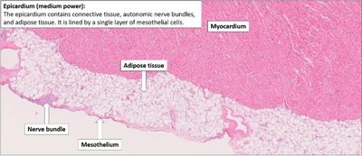
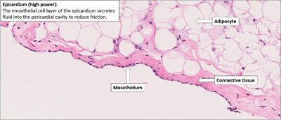
The epicardium is composed of simple squamous mesothelium and loose connective tissue containing blood vessels and nerves. It cushions the heart and reduces friction during movement by secreting serous fluid. The epicardium is continuous with the parietal pericardium at the base of large vessels.
Myocardium
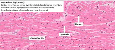
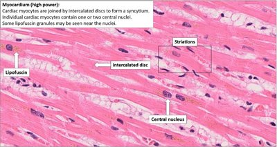
The myocardium is the thickest layer, consisting of spirally arranged cardiac muscle fibers. It is responsible for generating the force needed to pump blood. The left ventricular myocardium is about three times thicker than the right, reflecting its role in systemic circulation.
Cardiac myocytes are joined by intercalated discs, forming a functional syncytium.
Contains one or two central nuclei per cell and may show lipofuscin granules.
Endocardium
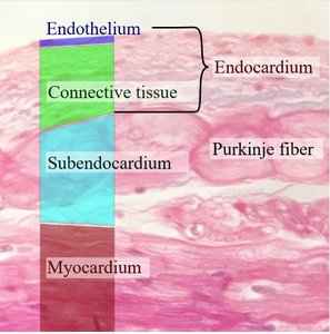
The endocardium lines the inner surface of the heart and is composed of three sublayers:
Endothelium: Simple squamous epithelium.
Subendothelial connective tissue: Supports the endothelium.
Subendocardial layer: Contains connective tissue and the heart's conduction system (Purkinje fibers).
Purkinje Fibers
Purkinje fibers are specialized cardiac muscle cells located in the subendocardial layer. They are large, pale-staining, and rich in glycogen, which makes them appear clear in H&E staining. Their primary function is to rapidly conduct electrical impulses from the AV node to the ventricular myocardium, ensuring coordinated ventricular contraction.
Cardiac Skeleton
The cardiac skeleton is a dense connective tissue framework surrounding the heart valves and the bases of the great vessels. It provides structural support, serves as an attachment site for the myocardium, and electrically isolates the atria from the ventricles, ensuring that conduction passes only through the AV node.
Blood Vessels
General Structure of Vessel Walls
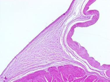
Blood vessels (except capillaries) share a common structural organization with three layers:
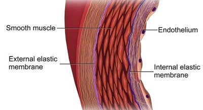
Tunica intima: Endothelium and subendothelial connective tissue; internal elastic lamina in arteries.
Tunica media: Helically arranged smooth muscle, elastic fibers, and other extracellular matrix components; external elastic lamina in arteries.
Tunica adventitia: Dense connective tissue, vasa vasorum, and vasomotor nerves; merges with surrounding tissue.
Elastic Arteries
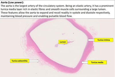
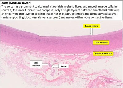
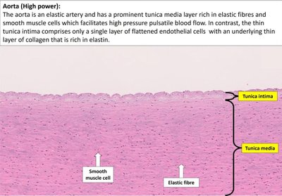
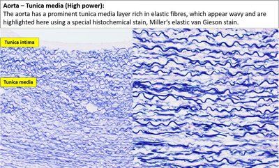
Elastic arteries (e.g., aorta) have a thick tunica media with alternating layers of elastic lamellae and smooth muscle. This structure allows them to withstand and dampen the pressure fluctuations of systole and diastole, maintaining continuous blood flow.
Tunica intima: Well-developed, may show folds postmortem.
Tunica media: Thickest layer, rich in elastic lamellae.
Tunica adventitia: Thinner than media, merges with connective tissue.
Vasa Vasorum
The vasa vasorum are small blood vessels that supply the outer layers of large arteries and veins, particularly in the tunica adventitia and outer tunica media. They provide nutrients and oxygen to vessel walls and remove metabolic wastes. Vasa vasorum are more abundant in veins due to lower oxygen content in venous blood.
Muscular Arteries
Muscular arteries regulate blood pressure by contracting or relaxing the smooth muscle in their tunica media. They have a prominent internal elastic lamina and up to 40 layers of smooth muscle cells. The tunica adventitia contains vasa vasorum, lymphatic capillaries, and nerves.
Arterioles
Arterioles are small vessels that control blood flow into capillary beds. Their media contains 1–2 layers of circular smooth muscle, acting as precapillary sphincters. Arterioles are major determinants of systemic blood pressure due to their muscle tone and ability to constrict or dilate.
Capillaries
Capillaries are the smallest blood vessels, consisting of a single layer of endothelial cells and a basement membrane. Their thin walls, large surface area, and slow blood flow optimize exchange of water, solutes, and macromolecules between blood and tissues. Only about 5% of blood volume is in capillaries at any time.
Pericytes: Support endothelial cells, regulate blood flow, maintain the blood-brain barrier, and contribute to tissue regeneration after injury.
Venules
Venules are transitional vessels between capillaries and veins. Postcapillary venules are primary sites for leukocyte extravasation during inflammation. Collecting and muscular venules have larger diameters and more smooth muscle.
Veins
Veins return blood to the heart under low pressure. Blood flow is aided by smooth muscle contraction, skeletal muscle compression, and the presence of valves (especially in limbs). Veins have thin walls, large lumens, and a thick tunica adventitia. Elastic fibers are present in large veins, and the internal elastic lamina may be seen in some cases.
Comparison of Arteries, Veins, and Capillaries
The following table summarizes the structural and functional differences among arteries, veins, and capillaries:
Arteries | Veins | Capillaries | |
|---|---|---|---|
Direction of Flow | Away from heart | Toward heart | Between arterioles & venules |
Pressure | High | Low | Very low |
Wall Thickness | Thick | Thin | Very thin (1 cell layer) |
Lumen | Narrow, round | Wide, irregular | Very narrow (RBCs pass single file) |
Tunica Media | Thick, smooth muscle & elastic fibers | Thin, few smooth muscle | Absent |
Tunica Adventitia | Relatively thin | Thickest layer | Absent |
Elastic Lamina | Prominent | Absent/inconspicuous | Absent |
Valves | Absent | Present (esp. limbs) | Absent |
Function | Distribute blood | Return blood, reservoir | Exchange of gases, nutrients, wastes |
End of discussion. Continue persevering, future RMT!
