 Back
The Cell Cycle and Cellular Reproduction: Structure, Regulation, and Clinical Relevance
Study Guide - Smart Notes
 Tailored notes based on your materials, expanded with key definitions, examples, and context.
The Cell Cycle
Overview of the Cell Cycle
The cell cycle is an orderly sequence of events that leads to cell growth, DNA replication, and cell division in eukaryotic cells. It ensures that genetic material is accurately duplicated and distributed to daughter cells.
Interphase: The longest phase, where the cell grows, performs normal functions, and prepares for division. Subdivided into G1, S, and G2 phases.
Mitotic (M) Phase: Includes mitosis (nuclear division) and cytokinesis (cytoplasmic division).
Checkpoints (G1, G2, M) regulate progression through the cycle, ensuring DNA integrity and proper chromosome alignment.
Phases of Interphase
G1 Phase: Cell recovers from previous division, grows, and synthesizes organelles and raw materials for DNA replication.
S Phase: DNA replication occurs, producing sister chromatids joined at the centromere.
G2 Phase: Final preparations for mitosis, including synthesis of proteins and microtubules.
Mitotic Stage
Mitosis: Division of the nucleus, distributing chromosomes to daughter nuclei.
Cytokinesis: Division of the cytoplasm, resulting in two genetically identical daughter cells.
Control of the Cell Cycle
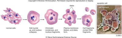
The cell cycle is regulated by internal (e.g., cyclins) and external signals (e.g., growth factors). Checkpoints ensure damaged DNA is repaired or the cell undergoes apoptosis (programmed cell death) if irreparable.
Cyclins: Proteins that regulate progression through the cell cycle.
p53 protein: Halts the cycle at G1 if DNA is damaged, initiates repair, or triggers apoptosis if repair fails.
The Eukaryotic Chromosome
Structure and Organization
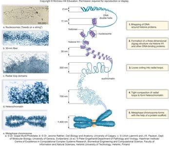
Eukaryotic chromosomes consist of a single DNA molecule associated with histone proteins, forming chromatin. Chromatin can be loosely packed (euchromatin, active) or highly compacted (heterochromatin, inactive).
Nucleosome: DNA wrapped around a core of eight histones, appearing as "beads on a string".
Further folding and coiling produce the condensed chromosome structure visible during mitosis.
Chromosome Number
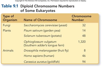
Each species has a characteristic diploid (2n) chromosome number. Humans have 46 chromosomes (23 pairs), with one set from each parent. Gametes (sperm and eggs) are haploid (n), containing one set of chromosomes.
Type of Organism | Name of Chromosome | Chromosome Number |
|---|---|---|
Fungi | Saccharomyces cerevisiae (yeast) | 32 |
Plants | Pisum sativum (garden pea) | 14 |
Plants | Solanum tuberosum (potato) | 48 |
Plants | Ophioglossum vulgatum (Southern adder's tongue fern) | 1,320 |
Animals | Drosophila melanogaster (fruit fly) | 8 |
Animals | Homo sapiens (human) | 46 |
Animals | Carassius auratus (goldfish) | 94 |
Mitosis and Cytokinesis
Phases of Mitosis
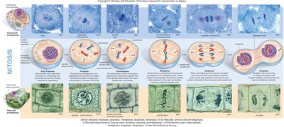
Mitosis is the process by which a eukaryotic cell separates its duplicated chromosomes into two identical sets. It consists of several stages:
Prophase: Chromatin condenses into visible chromosomes; spindle apparatus forms; nuclear envelope fragments.
Prometaphase: Kinetochores form at centromeres; spindle fibers attach to kinetochores.
Metaphase: Chromosomes align at the metaphase plate (cell equator).
Anaphase: Sister chromatids separate and move toward opposite poles.
Telophase: Nuclear envelopes reform around daughter chromosomes; chromosomes decondense.
Cytokinesis
Cytokinesis divides the cytoplasm, producing two daughter cells. The process differs between animal and plant cells:
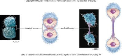
Animal cells: A cleavage furrow forms, pinching the cell in two via a contractile ring of actin filaments.
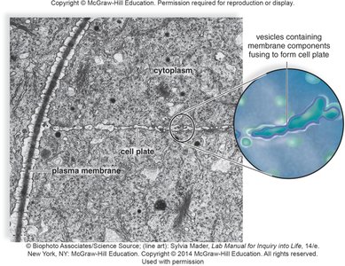
Plant cells: A cell plate forms from vesicles produced by the Golgi apparatus, eventually developing into a new cell wall.
Functions of Mitosis
Permits growth and repair in multicellular organisms.
In plants, meristematic tissue allows continuous division for growth.
In animals, stem cells enable tissue renewal and repair.
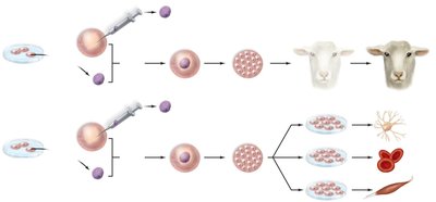
Therapeutic and reproductive cloning utilize mitosis for tissue generation and organismal cloning.
The Cell Cycle and Cancer
Characteristics of Cancer Cells
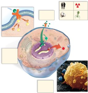
Cancer results from uncontrolled cell division due to mutations in genes regulating the cell cycle. Tumors can be benign (non-cancerous) or malignant (cancerous, invasive, and metastatic).
Lack differentiation: Cancer cells are non-specialized and can divide indefinitely.
Abnormal nuclei: Enlarged, irregular, and often with abnormal chromosome numbers.
Loss of apoptosis: Cancer cells evade programmed cell death.
Loss of contact inhibition: Cancer cells grow over one another, forming disorganized layers.
Metastasis: Cancer cells spread to distant tissues.
Angiogenesis: Formation of new blood vessels to supply the tumor.
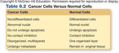
Cancer Cells | Normal Cells |
|---|---|
Nondifferentiated cells | Differentiated cells |
Abnormal nuclei | Normal nuclei |
Do not undergo apoptosis | Undergo apoptosis |
No contact inhibition | Contact inhibition |
Disorganized, multilayered | One organized layer |
Undergo metastasis | Remain in original tissue |
Genetic Basis of Cancer
Proto-oncogenes: Normally promote cell division; mutations convert them to oncogenes, causing uncontrolled division.
Tumor suppressor genes: Normally inhibit cell division and promote apoptosis; mutations inactivate these genes, removing growth inhibition.
p53 and RB genes: Key tumor suppressors; mutations are common in many cancers.
Causes of Cancer
Mutations can be caused by heredity, radiation, chemicals (pesticides, herbicides), and viruses.
Telomerase mutations allow cancer cells to maintain telomere length and divide indefinitely.
Prokaryotic Cell Division
Binary Fission
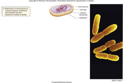
Prokaryotes (bacteria and archaea) divide by binary fission, a simpler process than mitosis. The single, circular chromosome replicates, and the cell splits into two genetically identical daughter cells.
Chromosome attaches to the plasma membrane and is duplicated.
Cell elongates, and new cell wall and membrane form between chromosomes.
No spindle apparatus is involved.
Comparison: Binary Fission vs. Mitosis
Both processes ensure genetic consistency in daughter cells.
Binary fission is typical of single-celled organisms; mitosis is essential for growth, repair, and development in multicellular eukaryotes.
Summary: The cell cycle is a tightly regulated process essential for growth, development, and tissue maintenance. Disruptions in cell cycle regulation can lead to cancer. Understanding the mechanisms of cell division in both eukaryotes and prokaryotes is fundamental to biology and medicine.
