 Back
BackThe Cell Cycle and Mitosis: Mechanisms of Eukaryotic Cell Division
Study Guide - Smart Notes
 Tailored notes based on your materials, expanded with key definitions, examples, and context.
The Cell Cycle and Mitosis
Introduction to Cell Division
Cell division is a fundamental process in all living organisms, enabling reproduction, growth, development, and tissue repair. In eukaryotes, this process is tightly regulated and occurs through a series of well-defined stages collectively known as the cell cycle. The most common form of cell division in somatic cells is mitosis, which ensures genetic continuity by producing two genetically identical daughter cells.
Why Do Cells Divide?
Unicellular Organisms: Cell division serves as a means of reproduction. In prokaryotes, this process is called binary fission.
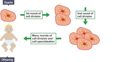
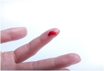
Multicellular Organisms: Cell division is essential for development from a single cell (zygote), organismal growth, and the repair of damaged tissues.
The Cell Cycle: Phases and Overview
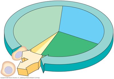
The cell cycle is the ordered sequence of events that a cell undergoes from its formation to its division into two daughter cells. It consists of two main phases: Interphase and the Mitotic (M) Phase.
Interphase: The cell grows, performs its normal functions, and duplicates its DNA. This phase accounts for about 90% of the cell cycle.
Mitotic (M) Phase: The cell divides its nucleus (mitosis) and cytoplasm (cytokinesis), accounting for about 10% of the cycle.
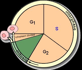
Subphases of Interphase
G1 Phase (First Gap): Cell grows and carries out normal metabolic functions.
S Phase (Synthesis): DNA is replicated, resulting in duplicated chromosomes.
G2 Phase (Second Gap): Cell continues to grow and prepares for division.
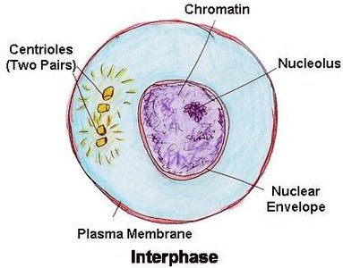
Chromatin and Chromosomes
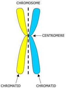
During interphase, DNA exists as chromatin, a loosely packed form that allows for gene expression and DNA replication. Prior to mitosis, chromatin condenses into visible chromosomes, each consisting of two identical sister chromatids joined at a region called the centromere.
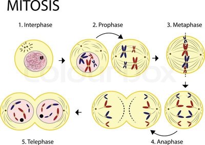

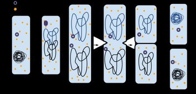
Mitosis: The Phases (PMAT)
Mitosis is divided into four main phases, often abbreviated as PMAT: Prophase, Metaphase, Anaphase, and Telophase. Each phase is characterized by distinct structural changes in the cell.

Prophase
Duplicated DNA condenses into chromosomes, each with two sister chromatids.
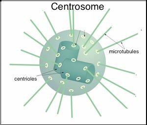
Centrosomes (organelles that organize microtubules) move to opposite poles and begin forming the mitotic spindle.
The nuclear envelope disintegrates, allowing spindle fibers to interact with chromosomes.
Metaphase
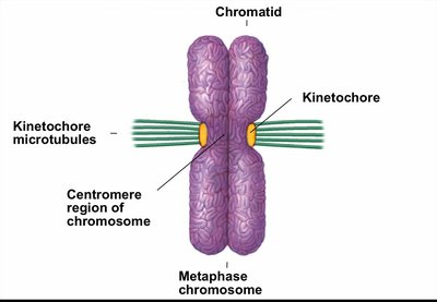
A protein structure called the kinetochore forms at the centromere of each chromosome.
Spindle fibers attach to kinetochores and align chromosomes along the metaphase plate (the cell's equator).

Anaphase
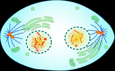
Sister chromatids are separated as spindle fibers shorten, pulling them toward opposite poles of the cell.
This movement is powered by the shortening of microtubules and the action of motor proteins.

Telophase
Nuclear envelopes reform around the two sets of chromosomes at each pole.
Chromosomes decondense back into chromatin.
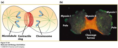
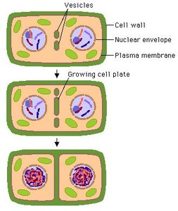
Cytokinesis: Division of the Cytoplasm
Cytokinesis is the process by which the cytoplasm divides, resulting in two separate daughter cells. The mechanism differs between animal and plant cells:
Animal Cells: A contractile ring of actin filaments forms a cleavage furrow that pinches the cell in two.
Plant Cells: Vesicles from the Golgi apparatus coalesce at the center of the cell to form a cell plate, which develops into a new cell wall separating the daughter cells.

Summary Table: Key Events of Mitosis
Phase | Main Events |
|---|---|
Prophase | Chromosomes condense, spindle forms, nuclear envelope breaks down |
Metaphase | Chromosomes align at metaphase plate, spindle fibers attach to kinetochores |
Anaphase | Sister chromatids separate and move to opposite poles |
Telophase | Nuclear envelopes reform, chromosomes decondense |
Cytokinesis | Cytoplasm divides, forming two daughter cells |
Key Terms and Concepts
Chromatin: The uncondensed form of DNA present during interphase.
Chromosome: A condensed DNA structure visible during mitosis, consisting of two sister chromatids joined at a centromere.
Sister Chromatids: Identical copies of a chromosome connected by a centromere.
Centromere: The region where sister chromatids are most closely attached.
Kinetochore: Protein complex at the centromere where spindle fibers attach.
Mitotic Spindle: Structure made of microtubules that segregates chromosomes during mitosis.
Cytokinesis: Division of the cytoplasm, distinct from nuclear division (mitosis).
Equations and Additional Information
Cell Cycle Duration: The relative duration of each phase can be represented as a proportion of the total cell cycle time.
Genetic Consistency: Mitosis ensures that each daughter cell receives an identical set of chromosomes (2n → 2n).
Equation for DNA content during the cell cycle:
Additional info: Errors in mitosis can lead to aneuploidy (abnormal chromosome number), which is associated with diseases such as cancer.
