 Back
The Cell Cycle and Mitosis: Structure, Regulation, and Cancer
Study Guide - Smart Notes
 Tailored notes based on your materials, expanded with key definitions, examples, and context.
The Cell Cycle
Overview of Cell Division
Cell division is a fundamental process in all living organisms, enabling growth, development, and tissue repair. In unicellular organisms, cell division reproduces the entire organism, while in multicellular organisms, it is essential for development from a fertilized cell, growth, and repair of tissues. Cell division is a key part of the cell cycle, which describes the life of a cell from its formation to its own division.
Mitosis produces two genetically identical daughter cells.
The genome is all the DNA in a cell. Prokaryotes have a single circular DNA molecule, while eukaryotes have multiple linear chromosomes.
Eukaryotic Chromosomes
Eukaryotic chromosomes are composed of chromatin, a complex of DNA and proteins. Each species has a characteristic number of chromosomes per cell. Somatic (body) cells are diploid (two sets of chromosomes), while gametes (sperm and egg) are haploid (one set).
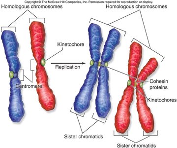
Chromosomes must be replicated before cell division.
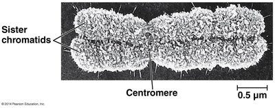
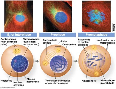
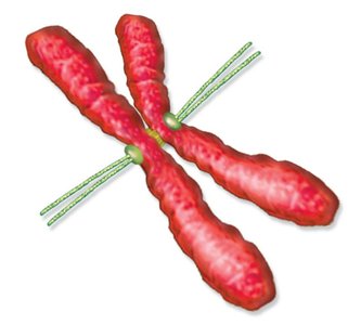
Replicated chromosomes are joined at the centromere and are called sister chromatids.
Cohesin proteins hold sister chromatids together.
Kinetochore proteins are attachment sites for spindle microtubules.
Chromosome Duplication and Division
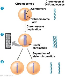
During the cell cycle, chromosomes are duplicated and then separated into daughter cells. The process ensures that each new cell receives an identical set of chromosomes.
After duplication, each chromosome consists of two sister chromatids.
During mitosis, sister chromatids separate and are distributed to two nuclei.
Phases of the Eukaryotic Cell Cycle
Main Phases
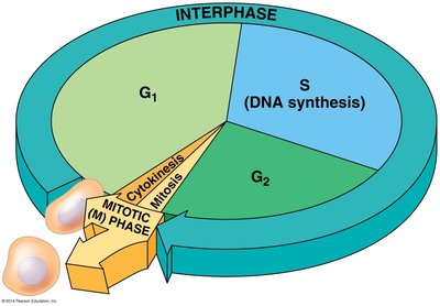
The eukaryotic cell cycle is divided into five main phases:
G1 (Gap 1)
S (Synthesis)
G2 (Gap 2)
M (Mitosis)
C (Cytokinesis)
Interphase (G1, S, G2) accounts for about 90% of the cell cycle, during which the cell grows and DNA is replicated (only during S phase).
Mitosis
Phases of Mitosis
Mitosis is the process of nuclear division and is divided into five phases:
Prophase
Prometaphase
Metaphase
Anaphase
Telophase
Each phase is characterized by specific events involving chromosomes, the spindle apparatus, and the nuclear envelope.
Prophase
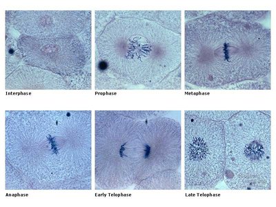
During prophase, chromosomes condense and become visible, the nucleoli disappear, and the mitotic spindle begins to form. In animal cells, centrosomes move to opposite poles and asters form around centrioles.
Prometaphase
The nuclear envelope fragments, chromosomes condense further, and kinetochores mature and attach to spindle microtubules. The spindle apparatus is completed.
Metaphase
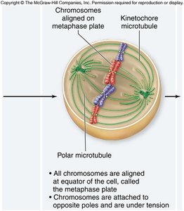
Chromosomes align at the metaphase plate, an imaginary plane equidistant from the spindle poles. Kinetochores attach chromosomes to spindle microtubules, and centromeres begin to divide.
Anaphase
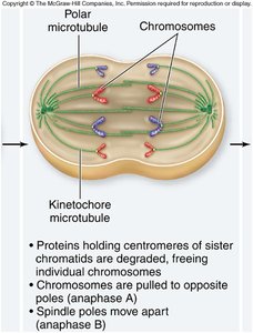
Cohesin proteins are degraded, allowing sister chromatids to separate. Microtubules pull chromatids toward opposite poles, and the spindle elongates the cell.
Telophase
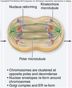
The spindle apparatus disassembles, nuclear envelopes reform around each set of chromosomes, chromosomes decondense, and nucleoli reappear.
Cytokinesis
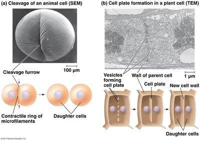
Cytokinesis is the division of the cytoplasm, resulting in two daughter cells. In animal cells, a cleavage furrow forms via actin filaments. In plant cells, a cell plate forms to separate the two nuclei.
Bacterial Cell Division
Binary Fission
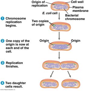
Bacteria divide by binary fission, a simpler process than mitosis. The single, circular chromosome is replicated, and each copy is segregated to opposite ends of the cell. A septum forms, dividing the cell into two daughter cells.
Regulation of the Cell Cycle
Cell Cycle Control System
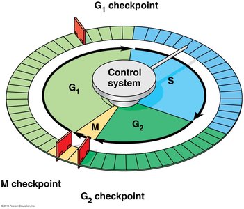
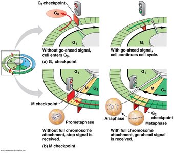
The cell cycle is regulated by a control system with checkpoints at critical stages. These checkpoints ensure that the cell only proceeds to the next phase when conditions are favorable.
G1/S checkpoint: Cell commits to division.
G2/M checkpoint: Cell commits to mitosis.
Spindle (M) checkpoint: Ensures all chromosomes are attached to the spindle before anaphase.
Chemical Control: Cyclins and Cdks
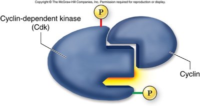
Cyclins are proteins whose concentrations fluctuate with the cell cycle. Cyclin-dependent kinases (Cdks) are enzymes activated by binding to cyclins, driving the cell through checkpoints by phosphorylating target proteins.
External Regulation
Growth factors: Chemicals or hormones that stimulate cell division (e.g., PDGF in wound healing).
Density-dependent inhibition: Crowded cells stop dividing.
Anchorage dependence: Cells must be attached to a substrate to divide.
Cancer and the Cell Cycle
Loss of Cell Cycle Control
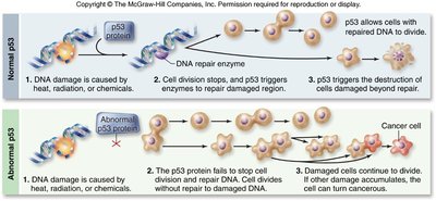
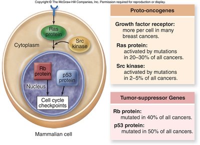
Cancer results from the failure of cell cycle control, leading to uncontrolled cell division and the formation of tumors. Cancer cells can divide indefinitely, invade other tissues, and metastasize to distant sites.
Tumor-suppressor genes (e.g., p53): Prevent cell division when DNA is damaged. Mutations in these genes can lead to cancer.
Proto-oncogenes: Encode proteins that promote cell division. When mutated, they become oncogenes and can drive cancer progression.
Tumor Types and Treatment
Benign tumors: Remain at the original site.
Malignant tumors: Invade surrounding tissues and can metastasize.
Treatment options include surgery, radiation, chemotherapy, and targeted therapies based on genetic mutations.
Summary Table: Key Terms and Concepts
Term | Definition |
|---|---|
Chromatin | Complex of DNA and protein in eukaryotic chromosomes |
Centromere | Region where sister chromatids are joined |
Cohesin | Protein complex holding sister chromatids together |
Kinetochore | Protein structure for spindle attachment |
Cyclin | Regulatory protein controlling cell cycle progression |
Cdk | Cyclin-dependent kinase, enzyme driving cell cycle |
p53 | Tumor-suppressor gene involved in DNA damage response |
Oncogene | Mutated proto-oncogene causing cancer |
