 Back
BackThe Cell Cycle and Mitosis: Structure, Function, and Regulation
Study Guide - Smart Notes
 Tailored notes based on your materials, expanded with key definitions, examples, and context.
The Cell Cycle
Overview of the Cell Cycle
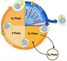
The cell cycle is the series of events that take place in a cell leading to its division and duplication. It is fundamental for growth, development, and maintenance in multicellular organisms. The cell cycle consists of interphase (G1, S, G2 phases) and the mitotic (M) phase, which includes mitosis and cytokinesis.
Interphase: The cell grows, performs its normal functions, and prepares for division by replicating its DNA.
Mitotic (M) Phase: The cell divides its copied DNA and cytoplasm to form two new cells.
Key Roles of Cell Division
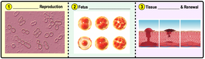
Cell division is essential for the continuity of life. It allows organisms to reproduce, grow, and repair tissues.
Reproduction: Unicellular organisms reproduce by cell division.
Growth and Development: Multicellular organisms develop from a single cell through repeated cell divisions.
Tissue Renewal: Cell division replaces cells that are lost due to normal wear and tear or injury.
Cell Division in Prokaryotes and Eukaryotes
Binary Fission in Prokaryotes
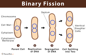
Prokaryotic cells, such as bacteria, divide by a process called binary fission. This process involves the replication of the single, circular chromosome, segregation of the DNA, and division of the cytoplasm, resulting in two genetically identical daughter cells.
Steps of Binary Fission:
Replication of DNA begins at the origin of replication.
The cell elongates as the DNA copies move apart.
The plasma membrane pinches inward, dividing the cell.
Cell Division in Eukaryotes
Eukaryotic cells divide by mitosis (for growth, repair, and asexual reproduction) or meiosis (for sexual reproduction, producing gametes). Mitosis ensures that each daughter cell receives an identical set of chromosomes.
Genetic Material Organization
Genome, Chromosomes, and Chromatin
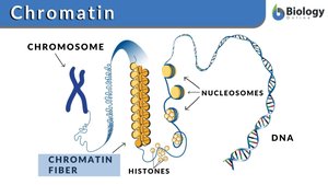
The genome is all the DNA in a cell. In eukaryotes, DNA is organized into multiple linear chromosomes, while prokaryotes typically have a single circular chromosome. Chromosomes are composed of chromatin, a complex of DNA and proteins (mainly histones) that help package the DNA efficiently.
Chromatin: DNA wrapped around histone proteins, forming nucleosomes and higher-order structures.
Chromosome: A condensed form of chromatin visible during cell division.
Structure of a Duplicated Chromosome
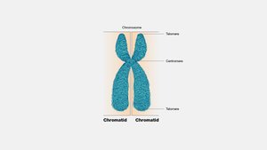
Before cell division, each chromosome is duplicated, forming two sister chromatids joined at a region called the centromere. The centromere is essential for the correct separation of chromatids during mitosis.
Sister Chromatids: Identical copies of a chromosome connected by a centromere.
Centromere: The region where the chromatids are most closely attached.
Phases of the Cell Cycle
Interphase
Interphase is the longest phase of the cell cycle, during which the cell grows, replicates its DNA, and prepares for division. It consists of three subphases:
G1 Phase (First Gap): Cell growth and normal metabolic roles.
S Phase (Synthesis): DNA replication and centrosome duplication.
G2 Phase (Second Gap): Further growth and preparation for mitosis.
Some cells may enter a non-dividing state called the G0 phase.
Mitosis: Division of the Nucleus
Overview of Mitosis
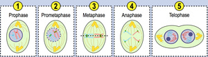
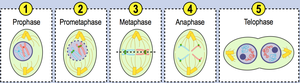
Mitosis is the process by which a eukaryotic cell separates its duplicated chromosomes into two identical sets, followed by division of the cytoplasm (cytokinesis). Mitosis is divided into several stages:
Prophase
Prometaphase
Metaphase
Anaphase
Telophase
Prophase
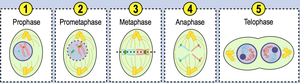
During prophase, chromatin condenses into visible chromosomes, each with two sister chromatids. The nucleolus disappears, and the mitotic spindle begins to form as centrosomes move to opposite poles of the cell.
Prometaphase
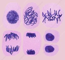
The nuclear envelope breaks down, allowing spindle microtubules to attach to chromosomes at the kinetochore, a protein structure on the centromere.
Metaphase
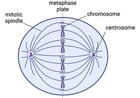
Chromosomes align at the cell's equator, known as the metaphase plate. The mitotic spindle is fully formed, and each chromosome is attached to spindle fibers from opposite poles.
Anaphase
Sister chromatids are separated and pulled toward opposite poles of the cell as spindle fibers shorten. This ensures each new cell will receive an identical set of chromosomes.
Telophase
Chromosomes arrive at the poles and begin to decondense back into chromatin. The nuclear envelope reforms around each set of chromosomes, resulting in two nuclei within the cell.
Cytokinesis
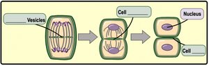
Cytokinesis is the division of the cytoplasm, which usually begins during telophase. In animal cells, a cleavage furrow forms to split the cell. In plant cells, vesicles from the Golgi apparatus form a cell plate that develops into a new cell wall.
Regulation of the Cell Cycle
Cell Cycle Control System
The cell cycle is regulated by a control system with checkpoints at G1, S, G2, and M phases. These checkpoints ensure that each phase is completed correctly before the next begins. If errors are detected, the cell can pause the cycle to repair damage or, if irreparable, initiate cell death (apoptosis).
G1 Checkpoint: Checks for DNA damage before replication.
S Checkpoint: Ensures proper DNA replication.
G2 Checkpoint: Confirms all components are ready for mitosis.
M (Metaphase) Checkpoint: Ensures chromosomes are properly attached to the spindle before separation.
Cancer and Cell Cycle Regulation
Cancer results from uncontrolled cell division due to mutations in genes that regulate the cell cycle. Two main types of genes are involved:
Proto-oncogenes: Promote normal cell division. When mutated, they become oncogenes and drive excessive cell division.
Tumor-suppressor genes: Inhibit cell division or promote apoptosis. Mutations in these genes remove growth restraints, contributing to cancer development.
Benign tumors do not spread, while malignant tumors can invade other tissues (metastasis).
Summary Table: Key Terms in the Cell Cycle
Term | Definition |
|---|---|
Genome | All the genetic material in a cell |
Chromosome | Condensed DNA molecule visible during cell division |
Chromatin | DNA-protein complex forming chromosomes |
Sister Chromatids | Identical copies of a duplicated chromosome |
Centromere | Region where sister chromatids are joined |
Kinetochore | Protein structure on centromere for spindle attachment |
Mitotic Spindle | Microtubule structure that separates chromosomes |
Cleavage Furrow | Indentation that begins cytokinesis in animal cells |
Cell Plate | Structure that forms during cytokinesis in plant cells |
