 Back
BackThe Cell Cycle, Mitosis, and Cancer: Regulation and Disease
Study Guide - Smart Notes
 Tailored notes based on your materials, expanded with key definitions, examples, and context.
The Cell Cycle and Its Regulation
Overview of the Cell Cycle
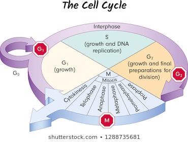
The cell cycle is the series of events that cells go through as they grow and divide. It consists of interphase (G1, S, G2) and the mitotic phase (mitosis and cytokinesis). Proper regulation ensures healthy growth, development, and tissue repair.
G1 phase: Cell grows and carries out normal functions.
S phase: DNA is replicated.
G2 phase: Cell prepares for division.
M phase (Mitosis): Division of the nucleus and cytoplasm.
G0 phase: Non-dividing state, often for specialized cells.
Checkpoints and Control Systems
Checkpoints are critical control mechanisms that ensure the cell cycle progresses only when conditions are favorable. The main checkpoints are at G1, G2, and M phases.
G1 checkpoint: Checks for cell size, nutrients, growth factors, and DNA damage.
G2 checkpoint: Ensures DNA replication is complete and undamaged.
M checkpoint: Verifies that all chromosomes are properly attached to the spindle before anaphase.
If a cell does not pass a checkpoint, it may enter G0 or activate repair or apoptosis mechanisms.
External and Internal Controls
Cell division is regulated by both internal and external signals:
Growth factors: Proteins that stimulate cell division.
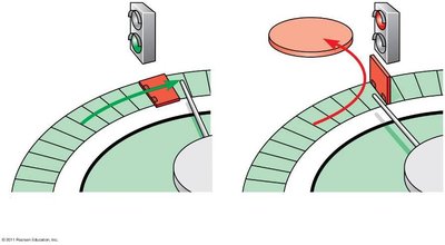
Density-dependent inhibition: Cells stop dividing when crowded.
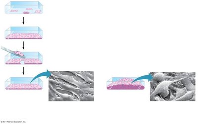
Anchorage dependence: Cells must be attached to a substrate to divide.
Mitosis: Stages and Identification
Phases of Mitosis
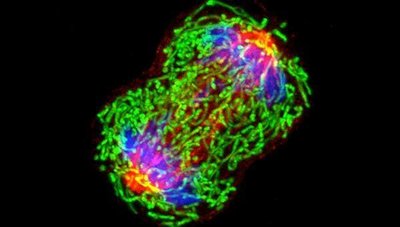
Mitosis is the process by which a eukaryotic cell separates its duplicated chromosomes into two identical nuclei. The stages are:
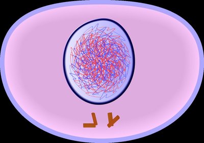
Prophase: Chromosomes condense, spindle forms, nuclear envelope breaks down.
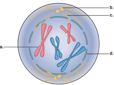
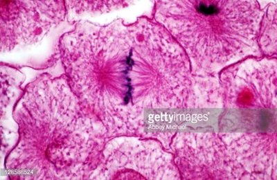
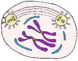
Metaphase: Chromosomes align at the cell's equator.
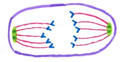
Anaphase: Sister chromatids separate and move to opposite poles.
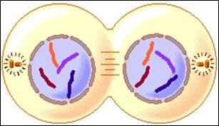
Telophase: Nuclear envelopes reform, chromosomes decondense.
Cytokinesis: Division of the cytoplasm, forming two daughter cells.
Interphase
Interphase is the longest phase, accounting for about 90% of the cell cycle. It includes G1, S, and G2 phases, during which the cell grows, replicates DNA, and prepares for division.
Cell Division Rates and Regulation
Variation in Cell Division Rates
Different cell types divide at different rates, depending on their function and developmental stage:
Skin cells in a fetus: Fastest division rate.
Skin cells in an adult: Fast division rate.
Liver cells: Slow division rate.
Nerve cells: Extremely slow or never divide (often in G0).
Cancer: Unregulated Cell Division
What is Cancer?
Cancer is a disease characterized by unregulated cell division. It arises when the normal controls of the cell cycle are disrupted, often due to mutations in genes that regulate cell growth and division.
More than 100 types, often named by tissue of origin.
Common feature: loss of cell cycle regulation.
Cell Division Driving Analogy
Cell cycle regulation can be compared to driving a car:
Accelerators: Proteins that promote cell division (e.g., growth factors, cyclins).
Brakes: Proteins that inhibit cell division (e.g., tumor suppressors).
Checkpoints act as safety checks, like a mechanic inspecting a car.

Genetic Basis of Cancer
Cancer is caused by mutations in two main types of genes:
Oncogenes: Mutated forms of genes (proto-oncogenes) that normally promote cell division. When mutated, they become overactive.
Tumor suppressor genes: Genes that normally inhibit cell division or promote apoptosis. When mutated, they lose function (e.g., p53).
Cancer usually requires mutations in multiple genes (6-9 or more).
The immune system also helps eliminate abnormal cells.
Loss of Growth Regulation in Cancer Cells
Cancer cells do not exhibit density-dependent inhibition or anchorage dependence, allowing them to grow uncontrollably and invade other tissues.

Tumor Types and Metastasis
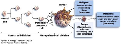
Tumors are classified based on their behavior:
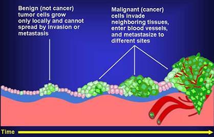
Benign tumor: Abnormal cells remain at the original site and do not invade other tissues (not cancer).
Malignant tumor: Invades surrounding tissues and can spread (cancerous).
Metastasis: Cancer cells spread to distant organs, forming secondary tumors.
Cancer Treatments
Several approaches are used to treat cancer, each targeting different aspects of cell division:
Surgery: Physical removal of tumors.
Radiation: Damages DNA in a localized area to prevent replication.
Chemotherapy: Drugs that inhibit DNA replication, cell division, or cell growth (affects all rapidly dividing cells).
Gene therapy: Experimental approaches to correct genetic defects.
Genetic Susceptibility and Heritability
Cancer is not directly inherited, but susceptibility can be. For example, mutations in the BRCA1 gene increase the risk of breast and ovarian cancer by impairing DNA repair mechanisms.
Contagious Cancers
Most cancers are not contagious, but there are rare exceptions:
HPV (Human Papilloma Virus): A sexually transmitted virus that increases cervical cancer risk by integrating its DNA into host tumor suppressor genes.
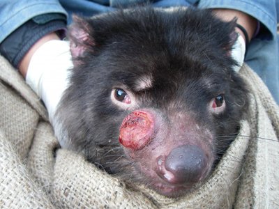
Devil Facial Tumor Disease (DFTD): A contagious cancer in Tasmanian devils, spread by biting. The cancer cells evade the immune system by lacking surface molecules.
Summary Table: Cell Cycle Checkpoints and Cancer
Checkpoint | Main Function | What Happens if Failed? |
|---|---|---|
G1 | Checks cell size, nutrients, DNA damage | Cell enters G0 or activates repair/apoptosis |
G2 | Checks DNA replication and damage | Cell cycle paused, repair or apoptosis |
M | Checks chromosome alignment | Cell cycle paused, repair or apoptosis |
Key Terms
Apoptosis: Programmed cell death, a mechanism to remove damaged cells.
Oncogene: A mutated gene that promotes uncontrolled cell division.
Tumor suppressor gene: A gene that inhibits cell division or induces apoptosis.
Metastasis: Spread of cancer cells to distant organs.
Density-dependent inhibition: Cells stop dividing when crowded.
Anchorage dependence: Cells require attachment to a substrate to divide.
Equations and Concepts
Cell cycle duration:
Mutation accumulation:
Additional info: This guide integrates textbook-level explanations and visual references to support understanding of cell cycle regulation, mitosis, and cancer biology, as covered in introductory college biology courses.
