 Back
BackThe Cell Cycle: Mitosis and Meiosis – Structure, Function, and Genetic Variability
Study Guide - Smart Notes
 Tailored notes based on your materials, expanded with key definitions, examples, and context.
The Cell Cycle: Mitosis and Meiosis
Overview of Chromosomes and Cell Types
The cell cycle is a fundamental process in biology, enabling growth, development, and reproduction in all living organisms. Chromosomes are discrete units of genetic material, and their structure and function are central to cell division. Cells in multicellular organisms are classified as germ-line cells (which give rise to gametes) and somatic cells (all other body cells).
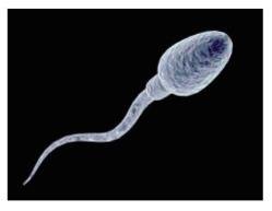
Germ-line cells: Produce gametes (egg and sperm).
Somatic cells: Include skin, brain, muscle cells, etc.
Cell division: Essential for growth, repair, and reproduction.
Levels of DNA Packaging
DNA is packaged in the cell nucleus in a highly organized manner. The structure varies from loosely packed chromatin to tightly packed chromosomes, especially during cell division.
Chromatin: DNA-protein complex forming eukaryotic chromosomes.
Chromosomes: Can be autosomes (homologous chromosomes) or sex chromosomes.
DNA packaging: Involves nucleosomes, histones, and higher-order structures.
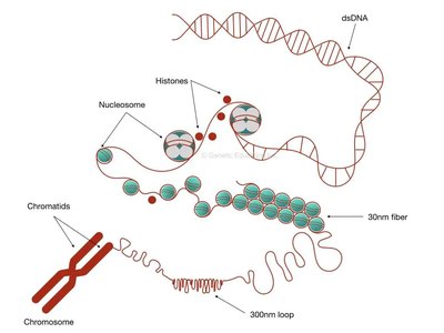
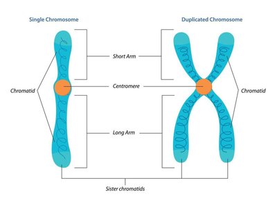
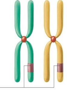
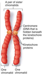
Sister Chromatids and Chromosome Duplication
Before cell division, DNA is replicated, resulting in two identical sister chromatids joined at the centromere. This process is crucial for accurate genetic material distribution.
Sister chromatids: Identical copies of a chromosome after DNA replication.
Centromere: Region where sister chromatids are joined.
Chromosome arms: Short and long arms distinguish chromosome structure.
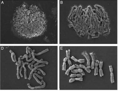
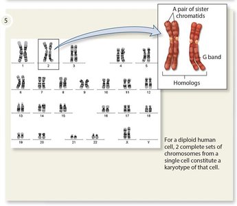
Chromosome Compaction and Visualization
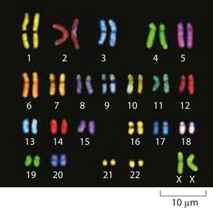
Chromosomes transition from loosely to tightly compacted forms during the cell cycle. Tightly compacted chromosomes are visible under a microscope and are used for karyotyping.
Karyotype: Photographic representation of chromosomes, showing number, size, and form.
Sets of Chromosomes and Ploidy
Organisms have characteristic numbers of chromosome sets. Human somatic cells are diploid (2N), while gametes are haploid (1N).
Ploidy: Number of chromosome sets (N).
Diploid (2N): Two sets of chromosomes (46 in humans).
Haploid (1N): One set of chromosomes (23 in humans).
Autosomes: Chromosomes 1–22.
Sex chromosomes: X and Y (XX = female, XY = male).

Homologous Chromosomes
In diploid cells, chromosomes exist in homologous pairs. Homologs carry the same genes but may have different alleles. Sex chromosomes (X and Y) are not homologous.
Homologs: Chromosome pairs with similar genes.
Alleles: Variants of a gene.
Sex chromosomes: X and Y carry different genes.
Types of Cell Division: Mitosis and Meiosis
Asexual vs Sexual Reproduction
Cell division is central to both asexual and sexual reproduction. Asexual reproduction produces genetically identical cells, while sexual reproduction generates genetic diversity.
Asexual reproduction: Mitosis; one parent; clones.
Sexual reproduction: Meiosis; two parents; genetically different offspring.

Mitosis: Process and Significance
Mitosis is the process by which a cell divides to produce two genetically identical daughter cells. It is essential for growth, development, and tissue repair in multicellular organisms.
Single-celled eukaryotes: Mitosis produces entire organisms.
Multicellular eukaryotes: Mitosis enables development, growth, and regeneration.
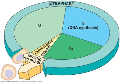
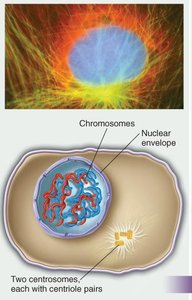
The Eukaryotic Cell Cycle
The cell cycle consists of interphase (G1, S, G2) and the mitotic (M) phase. Each stage has distinct events and checkpoints to ensure proper division.
G1 phase: Cell growth and decision to divide.
S phase: DNA replication; formation of sister chromatids.
G2 phase: Preparation for mitosis; error checking.
M phase: Mitosis and cytokinesis.
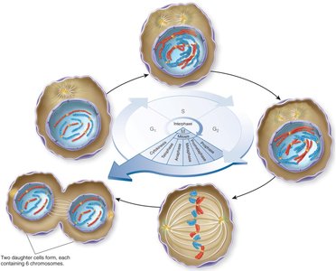


Mitosis: Phases and Checkpoints
Mitosis is divided into several phases, each with specific events. Checkpoints ensure accurate chromosome segregation.
Prophase: Chromosomes condense; nuclear envelope breaks down.
Prometaphase: Chromosomes finish condensing; spindle fibers attach.
Metaphase: Chromosomes align at metaphase plate; checkpoint ensures proper attachment.
Anaphase: Sister chromatids separate; move to opposite poles.
Telophase: Chromosomes decondense; nuclear envelope reforms.
Cytokinesis: Cytoplasm divides; two daughter cells form.

Cancer and the Cell Cycle
Cancer arises from uncontrolled cell division due to mutations in genes regulating the cell cycle. Tumors can be benign or malignant, with malignant tumors capable of metastasis.
Mutations: Permanent changes in DNA sequence.
p53 gene: Monitors DNA integrity; triggers repair or apoptosis.
Metastasis: Spread of cancer cells to other body parts.
Meiosis: Process and Genetic Variability
Role of Meiosis in Sexual Reproduction
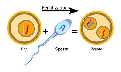
Meiosis is a specialized cell division process that reduces chromosome number by half, producing haploid gametes. Fertilization restores diploidy, forming a zygote.
Meiosis: Two rounds of cell division; produces four genetically unique haploid cells.
Fertilization: Union of egg and sperm to form a diploid zygote.
Genetic Variability in Sexual Reproduction
Sexual reproduction increases genetic variation through independent assortment, crossing over, and random fertilization. This variability is essential for evolutionary adaptation.
Independent assortment: Random distribution of homologous chromosomes.
Crossing over: Exchange of genetic material between homologs during prophase I.
Random fertilization: Any sperm can fuse with any egg.
Phases of Meiosis I and II
Meiosis consists of two sequential divisions: Meiosis I (separates homologous chromosomes) and Meiosis II (separates sister chromatids). Each phase has unique events contributing to genetic diversity.
Meiosis I: Prophase I (synapsis and crossing over), Metaphase I (independent assortment), Anaphase I (homolog separation), Telophase I (haploid cells).
Meiosis II: Prophase II, Metaphase II, Anaphase II (sister chromatid separation), Telophase II (haploid, genetically unique cells).
Comparison: Mitosis vs Meiosis
Mitosis and meiosis differ in the number of cell divisions, chromosome duplication rounds, and the genetic composition of daughter cells.
Feature | Mitosis | Meiosis |
|---|---|---|
Chromosome duplication | 1 round | 1 round |
Cell divisions | 1 | 2 |
Daughter cells produced | 2 | 4 |
Ploidy | Diploid (2N) | Haploid (1N) |
Genetic composition | Identical | Unique |
Summary
The cell cycle, mitosis, and meiosis are central to understanding cellular reproduction, genetic inheritance, and variability. These processes ensure continuity of life, adaptation, and evolution.
