 Back
BackThe Cell Cycle: Structure, Function, and Regulation
Study Guide - Smart Notes
 Tailored notes based on your materials, expanded with key definitions, examples, and context.
The Cell Cycle
Overview and Importance
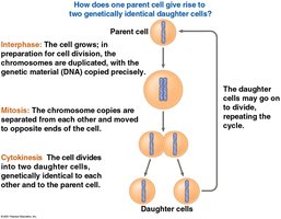
The cell cycle is the series of events that cells undergo as they grow and divide. It is fundamental to biological processes such as asexual reproduction, growth, development, and tissue renewal in multicellular organisms. Cell division is an integral part of the cell cycle, ensuring continuity of life and maintenance of organismal structure.
Asexual reproduction: In unicellular organisms, division of one cell reproduces the entire organism.
Growth and development: Multicellular eukaryotes depend on cell division for development from a fertilized egg and subsequent growth.
Tissue renewal: Cell division repairs and replaces damaged cells.
Genetic Identity in Cell Division
Most cell division, specifically mitosis, results in two daughter cells with identical genetic information. The exception is meiosis, which produces gametes (sperm and egg cells) with half the genetic material.
Mitosis: Produces genetically identical daughter cells.
Meiosis: Produces gametes with genetic variation (covered in Chapter 13).
Cellular Organization of Genetic Material
Chromosomes and Chromatin
All the DNA in a cell constitutes its genome. DNA molecules are packaged into chromosomes, which consist of chromatin—a complex of DNA and protein that condenses during cell division.
Somatic cells: Nonreproductive cells with two sets of chromosomes.
Gametes: Reproductive cells with half as many chromosomes as somatic cells.
Chromatin: DNA-protein complex that forms chromosomes.
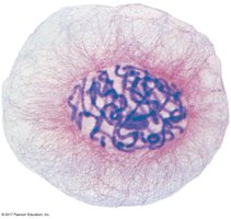
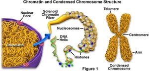
Chromosome Structure
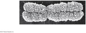
Chromosomes are composed of two sister chromatids joined at the centromere. Chromatin condenses to form visible chromosomes during cell division.
Sister chromatids: Joined copies of the original chromosome.
Centromere: The narrow region where chromatids are most closely attached.
Distribution of Chromosomes During Eukaryotic Cell Division
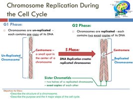
Chromosome Replication and Separation
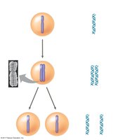
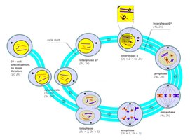
Before cell division, DNA is replicated and chromosomes condense. Each duplicated chromosome consists of two sister chromatids, which are separated during cell division and distributed to two nuclei. Once separated, chromatids are called chromosomes.
Chromosome duplication: Occurs during S phase of interphase.
Separation: Sister chromatids are pulled apart during mitosis.
The Cell Cycle Phases
Interphase and Mitotic Phase
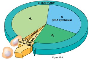
The cell cycle consists of interphase (cell growth and chromosome duplication) and the mitotic (M) phase (mitosis and cytokinesis). Interphase is subdivided into G1 (first gap), S (synthesis), and G2 (second gap) phases.
G1 phase: Cell growth and preparation for DNA replication.
S phase: DNA synthesis and chromosome duplication.
G2 phase: Preparation for mitosis.
Mitotic phase: Includes mitosis (nuclear division) and cytokinesis (cytoplasmic division).
Eukaryotic Cell Division: Mitosis and Cytokinesis
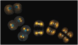
Mitosis
Mitosis is the division of the genetic material in the nucleus. It is followed by cytokinesis, which divides the cytoplasm. The process ensures that each daughter cell receives an identical set of chromosomes.
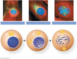
Prophase: Chromosomes condense, spindle forms.
Prometaphase: Nuclear envelope fragments, spindle fibers attach to kinetochores.
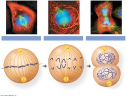
Metaphase: Chromosomes align at the metaphase plate.
Anaphase: Sister chromatids separate and move to opposite poles.
Telophase: Nuclear envelope reforms, chromosomes decondense.
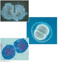
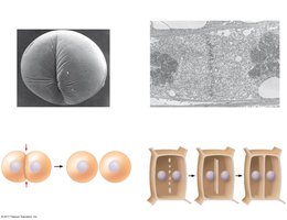
Cytokinesis
Cytokinesis is the division of the cytoplasm, resulting in two daughter cells. In animal cells, this occurs via cleavage; in plant cells, a cell plate forms.
Cleavage furrow: Contractile ring pinches the cell in two (animal cells).
Cell plate: Vesicles form a new cell wall (plant cells).
Regulation of the Eukaryotic Cell Cycle
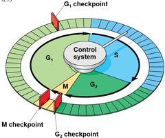
Cell Cycle Control System
The cell cycle is regulated by a molecular control system, functioning like a clock. This system is governed by internal and external controls and includes checkpoints where the cycle can be halted until conditions are favorable.
Checkpoints: G1, G2, and M checkpoints ensure proper progression.
Go-ahead signals: Required for the cell to proceed past checkpoints.
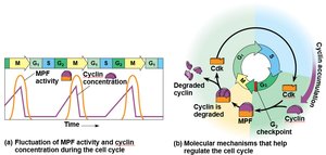
Cyclins and Cyclin-Dependent Kinases (Cdks)
Two types of regulatory proteins, cyclins and Cdks, control the cell cycle. The activity of Cdks fluctuates with the concentration of cyclins. MPF (maturation-promoting factor) is a cyclin-Cdk complex that triggers passage past the G2 checkpoint into the M phase.
Cyclins: Proteins whose levels rise and fall during the cell cycle.
Cdk: Enzymes activated by cyclins to regulate cell cycle events.
MPF: Cyclin-Cdk complex promoting mitosis.
G1 Checkpoint and G0 Phase
The G1 checkpoint is often the most critical. If a cell receives a go-ahead signal, it completes the cycle and divides. If not, it enters a nondividing state called G0.
G1 checkpoint: Determines whether the cell will proceed with division.
G0 phase: Nondividing, quiescent state.
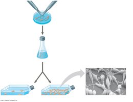
External Regulation: Growth Factors
External factors such as growth factors influence cell division. Platelet-derived growth factor (PDGF) stimulates fibroblast division, essential for wound healing.
Growth factors: Proteins released by cells to stimulate division in other cells.
PDGF: Produced by platelets, promotes connective tissue repair.
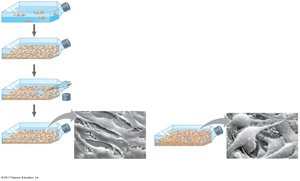
Anchorage and Density-Dependent Regulation
Normal cells require anchorage (a surface) and exhibit density-dependent inhibition (stop dividing when crowded). Cancer cells often lack these controls, leading to uncontrolled growth.
Anchorage dependence: Cells must be attached to a substrate to divide.
Density-dependent inhibition: Cells stop dividing when they form a single layer.
Cancer cells: Ignore these signals, leading to tumor formation.
Summary Table: Cell Cycle Phases and Key Events
Phase | Main Event | Key Structures |
|---|---|---|
G1 | Cell growth | Unreplicated chromosomes |
S | DNA replication | Duplicated chromosomes (sister chromatids) |
G2 | Preparation for mitosis | Replicated chromosomes |
Mitosis | Nuclear division | Mitotic spindle, chromosomes |
Cytokinesis | Cytoplasmic division | Cleavage furrow/cell plate |
Key Equations and Concepts
Chromosome number in somatic cells:
Chromosome number in gametes:
DNA replication:
Additional info:
Cell cycle regulation is crucial for preventing uncontrolled cell growth, which can lead to cancer.
Checkpoint failures are often associated with tumorigenesis.
