 Back
BackThe Cell Cycle: Structure, Function, and Regulation
Study Guide - Smart Notes
 Tailored notes based on your materials, expanded with key definitions, examples, and context.
The Cell Cycle
Key Roles of Cell Division
Cell division is fundamental to life, enabling organisms to reproduce, grow, and repair tissues. The continuity of life is maintained through the reproduction of cells, a process known as cell division.
Unicellular organisms: Division of one cell reproduces the entire organism.
Multicellular eukaryotes: Cell division is essential for development, growth, and repair.
Cell cycle: The life of a cell from its formation to its own division.
Genetic Information and Chromosome Distribution
Most cell division results in genetically identical daughter cells, except for meiosis, which produces gametes (sperm and egg cells) with half the chromosome number.
Genome: All the DNA in a cell; can be a single DNA molecule (prokaryotes) or multiple DNA molecules (eukaryotes).
Chromosomes: DNA molecules are packaged into chromosomes for efficient distribution during cell division.
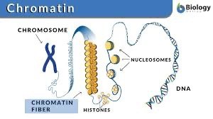
Chromatin Structure and DNA Packaging
Eukaryotic chromosomes are composed of chromatin, a complex of DNA and protein. Chromatin condenses during cell division to form visible chromosomes.
Nucleosomes: DNA wrapped around histone proteins, compacting DNA and regulating gene expression.
Chromatin vs. Chromosomes: DNA wraps around histones to form nucleosomes, which coil into chromatin, and further condense into chromosomes.
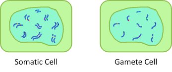
Chromosome Number and Cell Types
Each eukaryotic species has a characteristic number of chromosomes. Somatic cells have two sets of chromosomes, while gametes have half as many.
Somatic cells: Nonreproductive cells, diploid (2n).
Gametes: Reproductive cells, haploid (n).
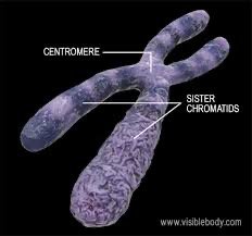
Chromosome Duplication and Sister Chromatids
Before cell division, DNA is replicated and chromosomes condense. Each duplicated chromosome consists of two sister chromatids joined at the centromere.
Sister chromatids: Identical copies of a chromosome, joined by cohesins.
Centromere: The region where chromatids are most closely attached.
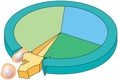
Phases of the Cell Cycle
Interphase and Mitotic Phase
The cell cycle consists of interphase (G1, S, G2) and the mitotic (M) phase. Interphase is the period of cell growth and DNA replication, while the M phase includes mitosis and cytokinesis.
G1 phase: Cell growth before DNA replication.
S phase: DNA replication and chromosomal protein duplication.
G2 phase: Preparation for cell division after DNA replication.
M phase: Mitosis (nuclear division) and cytokinesis (cytoplasmic division).
Mitosis: Steps and Mechanisms
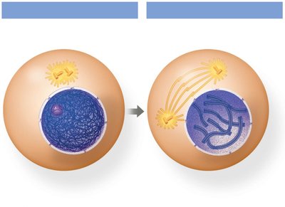
Prophase
During prophase, chromatin condenses into chromosomes, the nuclear envelope begins to break down, and the mitotic spindle forms.
Chromatin condenses: Chromosomes become visible, each consisting of two sister chromatids.
Mitotic spindle: Spindle fibers assemble to move chromosomes.
Centrosomes: Move to opposite poles, organizing spindle fibers.
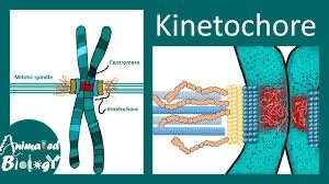
Prometaphase
The nuclear envelope breaks down completely, spindle fibers attach to chromosomes at kinetochores, and chromosomes begin moving.
Kinetochore: Protein structure at the centromere, attachment point for spindle microtubules.
Spindle apparatus: Fully developed, ensures proper chromosome movement.
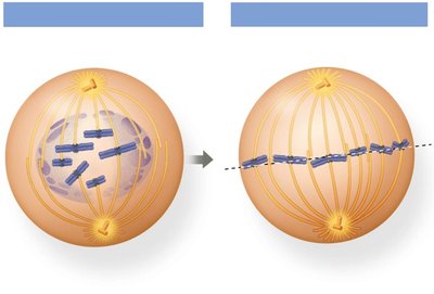
Metaphase
Chromosomes align at the metaphase plate, spindle fibers fully attach to kinetochores, and the spindle checkpoint ensures proper attachment and tension.
Metaphase plate: Imaginary line where chromosomes align.
Spindle checkpoint (SAC): Ensures all chromosomes are properly attached and tension is established.
Anaphase
Sister chromatids separate as cohesin is cleaved, chromosomes move to opposite poles, and the cell elongates.
Separase: Enzyme that cleaves cohesin, allowing chromatids to separate.
Spindle fibers: Shorten to pull chromosomes apart.
Equal distribution: Each pole receives an identical set of chromosomes.
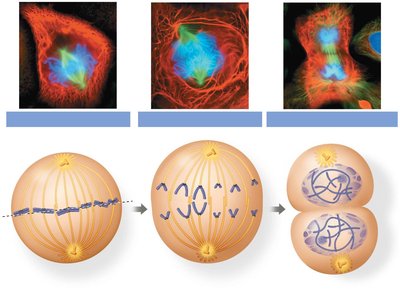
Telophase and Cytokinesis
Chromosomes arrive at opposite poles, decondense into chromatin, nuclear envelopes reform, and cytokinesis divides the cytoplasm.
Telophase: Chromosomes decondense, nucleoli reappear, nuclear envelope reforms.
Cytokinesis: In animal cells, cleavage furrow forms; in plant cells, a cell plate forms.

The Mitotic Spindle
Structure and Function
The mitotic spindle is composed of microtubules and controls chromosome movement during mitosis. It consists of three types of spindle fibers:
Kinetochore microtubules: Attach to chromosomes at kinetochores and pull chromatids apart.
Non-kinetochore microtubules: Do not attach to chromosomes; push against each other to elongate the cell.
Astral microtubules: Extend from centrosomes toward the cell membrane, anchoring the spindle.
Cell Cycle Control System
Regulation and Checkpoints
The cell cycle is regulated by internal and external controls, with checkpoints at G1, G2, and M phases. These checkpoints ensure proper cell size, DNA integrity, and chromosome attachment.
G1 checkpoint: Checks cell size, nutrients, DNA damage; decision point for division.
G2 checkpoint: Ensures DNA is fully replicated and undamaged.
M checkpoint: Ensures all chromosomes are attached to spindle fibers and aligned properly.
Cyclins and Cyclin-Dependent Kinases (Cdks)
Cyclins are proteins whose concentrations fluctuate during the cell cycle. Cyclin-dependent kinases (Cdks) are enzymes that phosphorylate target proteins, driving cell cycle progression. The cyclin-Cdk complex, known as MPF (maturation-promoting factor), triggers passage past the G2 checkpoint into mitosis.
MPF: Cyclin-Cdk complex that promotes mitosis.
Regulation: Cyclin synthesis and degradation control cell cycle timing.
External Factors and Cancer
Growth Factors and Cell Division
External factors such as growth factors stimulate cell division. Density-dependent inhibition and anchorage dependence regulate cell growth, preventing uncontrolled division.
Growth factors: Proteins released by cells to stimulate division in other cells.
Density-dependent inhibition: Crowded cells stop dividing.
Anchorage dependence: Cells must be attached to a surface to divide.
Loss of Cell Cycle Control in Cancer
Cancer cells do not respond to normal cell cycle controls, may produce their own growth factors, and can divide indefinitely. Tumors form when abnormal cells are not eliminated by the immune system. Benign tumors remain localized, while malignant tumors invade tissues and can metastasize.
Transformation: Process by which a normal cell becomes cancerous.
Metastasis: Spread of cancer cells to other parts of the body.
Summary Table: Cell Cycle Checkpoints
Checkpoint | Main Function | Key Questions |
|---|---|---|
G1 | Restriction point | Cell size, nutrients, DNA damage |
G2 | Readiness for mitosis | DNA fully replicated? Any damage? |
M | Spindle checkpoint | Chromosomes attached and aligned? |
Key Terms and Concepts
Cell cycle: The sequence of events in the life of a cell.
Mitosis: Division of the nucleus.
Cytokinesis: Division of the cytoplasm.
Chromatin: DNA-protein complex in eukaryotic cells.
Chromosome: Condensed form of chromatin during cell division.
Sister chromatids: Identical copies of a chromosome.
Centromere: Region joining sister chromatids.
Kinetochore: Protein structure for spindle attachment.
Spindle fibers: Microtubules involved in chromosome movement.
Cyclins/Cdks: Proteins and enzymes regulating cell cycle progression.
MPF: Cyclin-Cdk complex promoting mitosis.
Checkpoints: Control points ensuring proper cell cycle progression.
Growth factors: External signals stimulating cell division.
Cancer: Loss of cell cycle control, leading to uncontrolled division.
Additional info: The notes include expanded academic context and explanations for clarity and completeness, suitable for exam preparation.
