 Back
Chapter 6: The Cell - Structure, Function, and Microscopy
Study Guide - Smart Notes
 Tailored notes based on your materials, expanded with key definitions, examples, and context.
Chapter 6: The Cell - Structure, Function, and Microscopy
Introduction to the Study of Cells
The cell is the fundamental unit of life, and understanding its structure and function is central to biology. This chapter explores how cells are studied, why their size matters, the diversity of cell types, and the anatomy of both prokaryotic and eukaryotic cells.
Learning About Cells: Microscopy
Types of Microscopy
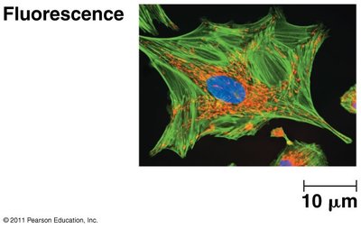
Light Microscopy: Uses visible light to observe cells. Includes brightfield, phase contrast, and fluorescence microscopy.
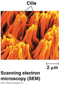
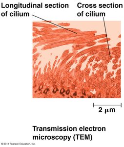
Electron Microscopy: Uses beams of electrons for much higher resolution. Includes scanning electron microscopy (SEM) and transmission electron microscopy (TEM).
Fluorescence microscopy allows visualization of specific cell components using fluorescent dyes or proteins.
SEM provides detailed surface images, while TEM reveals internal structures.
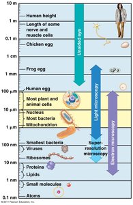
Scale in Microscopy: Different microscopes are suited for different size ranges, from atoms to whole cells.
Cell Size and Its Importance
Surface Area to Volume Ratio
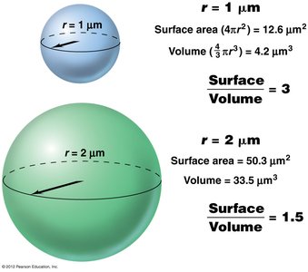
Cell size is limited by the surface area to volume ratio, which affects the efficiency of material exchange with the environment. As a cell grows, its volume increases faster than its surface area, limiting size.
Formula for Surface Area of a Sphere:
Formula for Volume of a Sphere:
Types of Cells: Prokaryotic vs. Eukaryotic
Key Differences
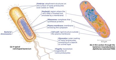
Prokaryotic Cells: Lack a nucleus and membrane-bound organelles. DNA is in the nucleoid region. Examples: Bacteria and Archaea.
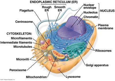
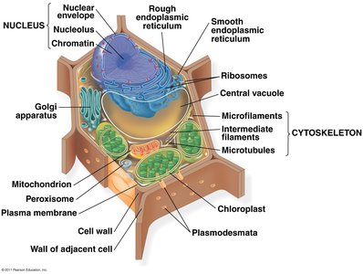
Eukaryotic Cells: Have a true nucleus and various membrane-bound organelles. Examples: Plants, animals, fungi, protists.
Cytosol refers to the fluid portion of the cytoplasm, while cytoplasm includes the cytosol and organelles (except the nucleus).
Cell Anatomy: Structure and Function of Organelles
Nucleus: The Cell’s Genetic Library
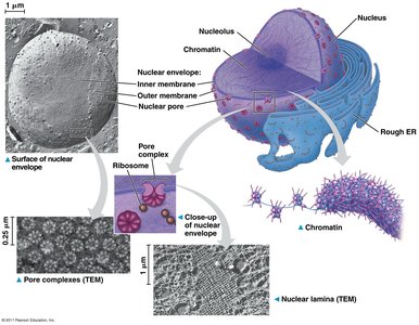
The nucleus stores genetic information and coordinates cell activities such as growth and reproduction. It is surrounded by a double membrane (nuclear envelope) and contains chromatin and the nucleolus.
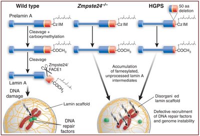
Nuclear Matrix Protein Defect: Progeria
Mutations in nuclear matrix proteins, such as lamin A, can cause diseases like progeria, a premature aging disorder. Lamin A is crucial for nuclear structure and DNA repair.
Ribosomes: The Cell’s Protein Factories
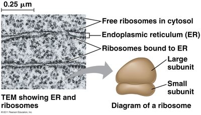
Ribosomes are complexes of RNA and protein that synthesize proteins. They can be free in the cytosol or bound to the endoplasmic reticulum (ER). Cells with high rates of protein secretion have many bound ribosomes.
The Endomembrane System
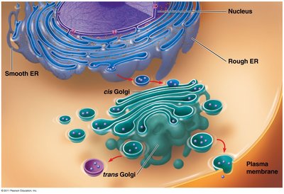
This system includes the nuclear envelope, ER, Golgi apparatus, lysosomes, vacuoles, and plasma membrane. It is essential for protein trafficking, metabolism, and cellular compartmentalization.
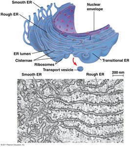
Endoplasmic Reticulum (ER): The Cell’s Biosynthetic Factory
Rough ER: Studded with ribosomes; synthesizes proteins for secretion or membrane insertion.
Smooth ER: Lacks ribosomes; involved in lipid synthesis, detoxification, and calcium storage. Abundant in liver cells.
Golgi Apparatus: The Cell’s Shipping & Receiving Center
The Golgi apparatus modifies, sorts, and packages proteins and lipids for delivery to various destinations.
Lysosomes: The Cell’s Digestive Center
Lysosomes contain hydrolytic enzymes for intracellular digestion. They break down macromolecules, old organelles, and pathogens. Lysosomal storage diseases, such as Gaucher disease, result from enzyme deficiencies.
Vacuoles: Storage and More
Plant Cells: Central vacuole stores ions, pigments, and nutrients; maintains turgor pressure.
Protists: Contractile vacuoles regulate water balance.
Mitochondria: The Cell’s Powerhouse
Mitochondria are the sites of cellular respiration, converting glucose and oxygen into ATP, the cell’s energy currency.
Chloroplasts: The Site of Photosynthesis
Chloroplasts, found in plant cells, capture light energy to synthesize glucose during photosynthesis. They are part of the plastid family, which also includes chromoplasts (pigment storage) and amyloplasts (starch storage).
Peroxisomes: The Cell’s Site of Oxidation
Peroxisomes contain enzymes that generate and degrade hydrogen peroxide, catabolize lipids, and convert fats to sugars (glyoxysomes in plants).
The Cytoskeleton: Support and Motility
Components of the Cytoskeleton
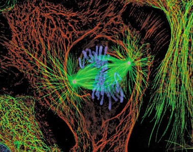
Microtubules: Hollow rods that maintain cell shape, facilitate organelle movement, and form the mitotic spindle.
Microfilaments: Thin filaments of actin involved in cell movement and muscle contraction.
Intermediate Filaments: Provide mechanical support; include keratin and lamin proteins.
Centrosomes and Basal Bodies
Centrosomes are microtubule-organizing centers in animal cells. Basal bodies anchor cilia and flagella.
Microtubules in Action: Cilia and Flagella
Cilia and flagella are motile structures powered by microtubules. Cilia move substances across cell surfaces; flagella propel cells.
Microfilaments in Action
Microfilaments interact with myosin for muscle contraction and cell movement.
Intermediate Filaments
Intermediate filaments provide tensile strength and structural integrity. Mutations can cause diseases affecting skin and other tissues.
Cell Surface and Interactions
Cell Walls
Plant Cells: Cellulose-based cell walls provide rigidity.
Fungi: Chitin-based cell walls.
The Extracellular Matrix (ECM)
The ECM is composed mainly of glycoproteins and provides structural support, regulates cell behavior, and mediates cell signaling.
Cell Junctions
Cell junctions connect cells and facilitate communication. Types include tight junctions, desmosomes, and gap junctions in animal cells; plasmodesmata in plant cells.
Summary Table: Major Eukaryotic Cell Structures and Functions
Organelle/Structure | Main Function |
|---|---|
Nucleus | Stores genetic material, coordinates cell activities |
Ribosome | Protein synthesis |
Endoplasmic Reticulum (Rough/Smooth) | Protein and lipid synthesis, detoxification |
Golgi Apparatus | Modification, sorting, and packaging of proteins/lipids |
Lysosome | Intracellular digestion |
Vacuole | Storage, waste disposal, water balance |
Mitochondrion | ATP production via cellular respiration |
Chloroplast | Photosynthesis (plants/algae) |
Peroxisome | Oxidation of fatty acids, detoxification |
Cytoskeleton | Structural support, movement |
Cell Wall | Protection, structural support (plants, fungi, some protists) |
Extracellular Matrix | Cell signaling, adhesion, support (animals) |
Cell Junctions | Cell-cell communication and adhesion |
