 Back
BackThe Cellular Basis of Reproduction and Inheritance
Study Guide - Smart Notes
 Tailored notes based on your materials, expanded with key definitions, examples, and context.
Chapter 8: The Cellular Basis of Reproduction and Inheritance
Introduction to Cell Division and Cancer
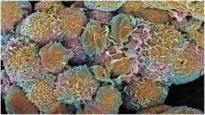
Cell division is a fundamental process in all living organisms, essential for growth, development, and maintenance. While normal cell division is tightly regulated, mutations can disrupt this control, leading to cancer. Cancer cells divide uncontrollably, invade other tissues, and can ultimately be fatal.
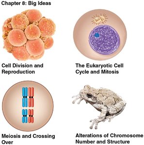
Big Ideas of Chapter 8
Cell Division and Reproduction
The Eukaryotic Cell Cycle and Mitosis
Meiosis and Crossing Over
Alterations of Chromosome Number and Structure
Cell Division and Reproduction
Roles of Cell Division
Cell division is central to the reproduction of cells and organisms. It ensures continuity of life by producing new cells from preexisting ones. There are two main types of reproduction:
Asexual reproduction: Offspring are genetic clones of the parent (e.g., binary fission in prokaryotes).
Sexual reproduction: Offspring are genetically diverse due to the combination of genetic material from two parents.
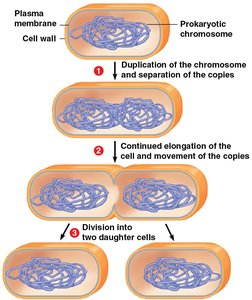
Prokaryotic Cell Division: Binary Fission
Prokaryotes reproduce asexually by binary fission, a process in which the cell divides in half to produce two genetically identical daughter cells. The steps include:
Duplication of the single, circular chromosome.
Separation of chromosome copies as the cell elongates.
Division of the parent cell into two daughter cells by inward growth of the plasma membrane and cell wall.
The Eukaryotic Cell Cycle and Mitosis
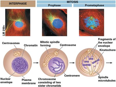
Eukaryotic Chromosomes and Chromatin
Eukaryotic cells contain multiple, linear chromosomes located in the nucleus. Each chromosome consists of a long DNA molecule and associated proteins. Chromosomes are only visible during cell division; otherwise, they exist as loosely packed chromatin.
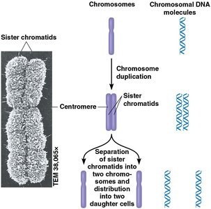
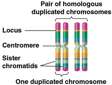
Chromosome Duplication and Sister Chromatids
Before a eukaryotic cell divides, each chromosome is duplicated, forming two sister chromatids joined at a region called the centromere. During cell division, sister chromatids separate, ensuring each daughter cell receives an identical set of chromosomes.
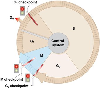
The Cell Cycle: Phases and Checkpoints
The cell cycle is an ordered sequence of events from the formation of a cell to its own division. It consists of:
Interphase: Includes G1 (first gap), S (DNA synthesis), and G2 (second gap) phases.
Mitotic (M) phase: Includes mitosis (nuclear division) and cytokinesis (cytoplasmic division).

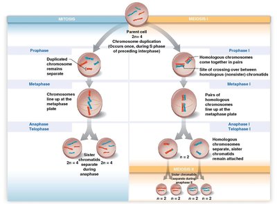
Mitosis: Distribution of Chromosomes
Mitosis is the process by which duplicated chromosomes are equally distributed into two daughter nuclei. It involves several stages:
Prophase: Chromosomes condense, spindle forms.
Prometaphase: Nuclear envelope fragments, spindle microtubules attach to kinetochores.
Metaphase: Chromosomes align at the metaphase plate.
Anaphase: Sister chromatids separate and move to opposite poles.
Telophase: Nuclear envelopes reform around chromosomes.
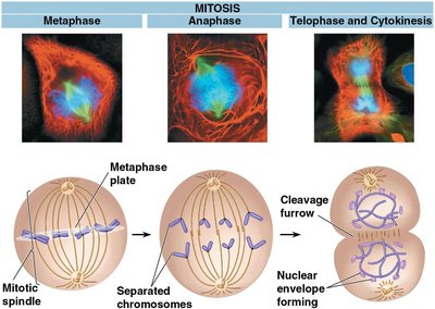
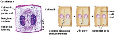
Cytokinesis: Animal vs. Plant Cells
Cytokinesis is the division of the cytoplasm, overlapping with the end of mitosis. The process differs between animal and plant cells:
Animal cells: A cleavage furrow forms, pinching the cell in two.
Plant cells: A cell plate forms, eventually developing into a new cell wall that separates the two daughter cells.

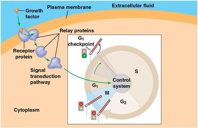
Regulation of the Cell Cycle
The cell cycle is regulated by a set of proteins that control progression through critical checkpoints (G1, G2, and M). External signals, such as growth factors, are often required for a cell to proceed through the cycle and divide.
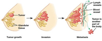
Cancer and Cell Division
Cancer results from the loss of cell cycle control, leading to excessive cell division and the formation of tumors. Malignant tumors can invade other tissues (metastasis). Treatments like radiation and chemotherapy target rapidly dividing cells.
Meiosis and Crossing Over
Homologous Chromosomes
Somatic cells contain pairs of homologous chromosomes, each carrying genes for the same traits at the same loci. Humans have 46 chromosomes (23 pairs). Not all chromosomes are fully homologous (e.g., sex chromosomes).
Haploid and Diploid Cells
Diploid cells (2n) have two sets of chromosomes, while haploid cells (n), such as gametes (egg and sperm), have one set. Sexual life cycles alternate between haploid and diploid stages.
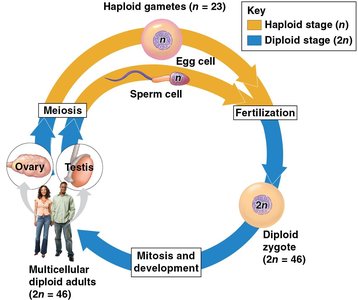
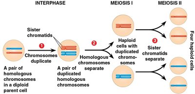
Meiosis: Reduction Division
Meiosis reduces the chromosome number from diploid to haploid, producing four genetically unique gametes. It involves two consecutive divisions:
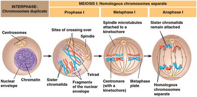
Meiosis I: Homologous chromosomes pair and separate, crossing over occurs.
Meiosis II: Sister chromatids separate, similar to mitosis.
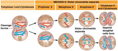
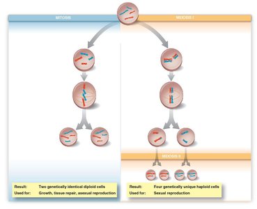
Comparing Mitosis and Meiosis
Both processes begin with chromosome duplication in diploid cells, but their outcomes differ:
Mitosis: Two genetically identical diploid cells (for growth, repair, asexual reproduction).
Meiosis: Four genetically unique haploid cells (for sexual reproduction).
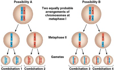
Genetic Variation: Independent Assortment and Crossing Over
Genetic diversity arises from:
Independent orientation: Random arrangement of homologous pairs during metaphase I leads to many possible chromosome combinations in gametes.
Random fertilization: Any sperm can fertilize any egg, further increasing variation.
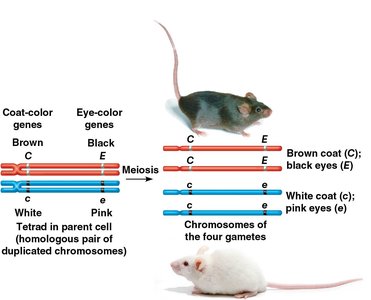
Crossing over: Exchange of genetic material between nonsister chromatids during prophase I creates new allele combinations.
Alterations of Chromosome Number and Structure
Nondisjunction and Chromosome Number Abnormalities
Nondisjunction is the failure of chromosomes or chromatids to separate properly during meiosis, resulting in gametes with abnormal chromosome numbers. This can lead to disorders such as Down syndrome (trisomy 21).
Karyotypes
A karyotype is a photographic inventory of an individual's chromosomes, arranged in pairs. It is used to detect chromosomal abnormalities.
Down Syndrome and Sex Chromosome Abnormalities
Trisomy 21 (Down syndrome) is caused by an extra copy of chromosome 21. Abnormal numbers of sex chromosomes (e.g., XXY, XO) can also occur, leading to various syndromes.
Sex Chromosomes | Syndrome | Origin of Nondisjunction | Symptoms |
|---|---|---|---|
XXY | Klinefelter syndrome (male) | Meiosis in egg or sperm formation | Sterile; underdeveloped testes; secondary female characteristics |
XYY | None (normal male) | Meiosis in sperm formation | None |
XXX | None (normal female) | Meiosis in egg or sperm formation | Slightly taller than average |
XO | Turner syndrome (female) | Meiosis in egg or sperm formation | Sterile; immature sex organs |
Polyploidy and Evolution
Errors in cell division can result in polyploid organisms (with extra sets of chromosomes), which is an important mechanism in the evolution of new species, especially in plants.
Alterations of Chromosome Structure
Chromosome breakage can lead to structural changes such as deletions, duplications, inversions, and translocations. These alterations can cause genetic disorders or contribute to cancer if they occur in somatic cells.
Example: Reciprocal translocation involves the exchange of segments between nonhomologous chromosomes, which is different from crossing over (exchange between homologous chromosomes).
