 Back
BackThe Chromosomal Basis of Inheritance
Study Guide - Smart Notes
 Tailored notes based on your materials, expanded with key definitions, examples, and context.
Chromosomes and Inheritance
Historical Foundations
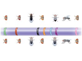
The concept that genes are located on chromosomes provided a physical basis for Mendel’s laws of inheritance. Thomas Hunt Morgan’s work with Drosophila melanogaster (fruit flies) demonstrated that genes occupy specific loci on chromosomes, and that the behavior of chromosomes during meiosis explains patterns of inheritance.
Gene: A unit of heredity located on a chromosome.
Locus (plural: loci): The specific location of a gene on a chromosome.
Mendel’s Laws: Law of Segregation and Law of Independent Assortment are explained by chromosomal behavior during meiosis.
Chromosomal Basis of Gender
Sex Chromosomes in Humans
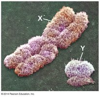
In humans and other mammals, sex is determined by the combination of sex chromosomes inherited. The X chromosome is larger and contains more genes than the Y chromosome.
XX: Female
XY: Male
Only the ends of the Y chromosome are homologous with the X chromosome.
X vs. Y Chromosomes and Sex-Linked Genes
SRY gene: Located on the Y chromosome, determines maleness.
Sex-linked gene: A gene located on either sex chromosome.
X-linked genes: Genes on the X chromosome; many are unrelated to sex determination.
Y-linked genes: Genes on the Y chromosome; relatively few in number.
Inheritance of X-Linked Genes
Patterns of X-Linked Inheritance
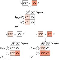
X-linked genes exhibit unique inheritance patterns due to the difference in X chromosome number between males and females.
Females (XX) require two copies of a recessive allele to express an X-linked trait.
Males (XY) require only one copy of a recessive allele to express the trait.
X-linked recessive disorders are more common in males (e.g., color blindness, Duchenne muscular dystrophy, hemophilia).
X Inactivation in Female Mammals
Barr Bodies and Genetic Mosaics
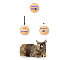
In female mammals, one X chromosome in each cell is randomly inactivated during embryonic development, forming a Barr body. This leads to mosaic expression of X-linked genes in heterozygous females.
Barr body: The condensed, inactive X chromosome in a female cell.
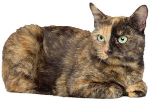
Females heterozygous for X-linked genes can show mosaic phenotypes (e.g., tortoiseshell cats).
Linked Genes and Genetic Recombination
Linked Genes
Genes located close together on the same chromosome tend to be inherited together and are called linked genes. However, linkage is not absolute due to recombination.
Linked genes do not assort independently.
Parental phenotypes are more common than recombinant phenotypes in offspring of linked genes.
Genetic Recombination and Crossing Over
Genetic recombination produces offspring with combinations of traits different from either parent. Crossing over during meiosis can break the linkage between genes, resulting in recombinant chromosomes.
Parental types: Offspring with phenotypes matching one of the parents.
Recombinant types: Offspring with new combinations of traits.
A 50% recombination frequency indicates genes are on different chromosomes or far apart on the same chromosome.
Mapping Genes Using Recombination Frequencies
Alfred Sturtevant developed genetic maps based on recombination frequencies. The farther apart two genes are, the higher the probability of a crossover event between them.
Linkage map: A genetic map based on recombination frequencies.
Map unit (centimorgan): Represents a 1% recombination frequency.
Genes far apart on the same chromosome can appear genetically unlinked.
Alterations of Chromosome Number and Structure
Aneuploidy and Polyploidy
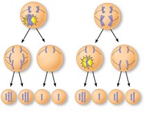
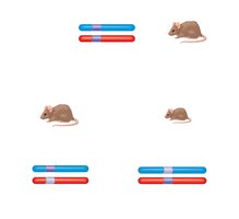
Errors in meiosis, such as nondisjunction, can result in abnormal chromosome numbers (aneuploidy) or extra sets of chromosomes (polyploidy).
Nondisjunction: Failure of homologous chromosomes or sister chromatids to separate properly during meiosis.
Aneuploidy: Abnormal number of a particular chromosome (e.g., monosomy, trisomy).
Polyploidy: More than two complete sets of chromosomes (common in plants).
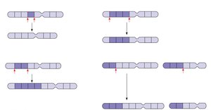
Alterations of Chromosome Structure
Chromosome breakage can lead to structural changes:
Deletion: Loss of a chromosomal segment.
Duplication: Repetition of a segment.
Inversion: Reversal of a segment within a chromosome.
Translocation: Movement of a segment to a nonhomologous chromosome.
Human Disorders Due to Chromosomal Alterations
Examples of Aneuploidy
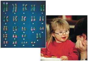
Down syndrome (Trisomy 21): Caused by three copies of chromosome 21; risk increases with maternal age.
Klinefelter syndrome (XXY): Extra X chromosome in males.
Turner syndrome (X0): Monosomy X in females; only known viable monosomy in humans.
Disorders from Chromosomal Structural Changes
Cri du chat syndrome: Deletion on chromosome 5; severe intellectual disability and characteristic cry.
Chronic myelogenous leukemia (CML): Caused by translocation between chromosomes 9 and 22 (Philadelphia chromosome).
Exceptions to Standard Mendelian Inheritance
Genomic Imprinting
Genomic imprinting is the phenomenon where the expression of an allele depends on whether it is inherited from the mother or the father. This is often due to methylation of DNA.
Imprinting affects only a small fraction of genes, but is critical for embryonic development.
Inheritance of Organelle Genes
Genes located in mitochondria and chloroplasts (extranuclear genes) are inherited maternally, as the zygote’s cytoplasm comes from the egg. Mutations in mitochondrial DNA can cause diseases affecting energy production.
Mitochondrial myopathy and Leber’s hereditary optic neuropathy are examples of disorders caused by mutations in mitochondrial genes.
