 Back
BackThe Chromosomal Basis of Inheritance: Study Notes
Study Guide - Smart Notes
 Tailored notes based on your materials, expanded with key definitions, examples, and context.
The Chromosomal Basis of Inheritance
Introduction
The chromosomal basis of inheritance explains how genes are transmitted from parents to offspring through chromosomes. This concept integrates Mendelian genetics with cytological observations of chromosomes during meiosis, providing a physical basis for the inheritance patterns first described by Gregor Mendel.
Chromosome Theory of Inheritance
Historical Foundations
Mendel's Gene Idea (1860s): Mendel proposed that genes are discrete units of inheritance, but the cellular basis was unknown at the time.
Discovery of Mitosis (1875) and Meiosis (1890): Observations revealed that chromosomes exist in pairs and segregate during cell division, paralleling Mendel's laws.
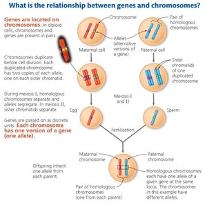
Chromosome Theory (1902): Genes are located on chromosomes at specific loci. Chromosomes undergo segregation and independent assortment during meiosis, explaining Mendel's observations.
Key Principles
Genes are located on chromosomes and are inherited as units.
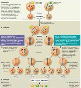
Chromosomes segregate during meiosis I (anaphase I), separating homologous pairs.
Independent assortment occurs during metaphase I, where chromosome pairs align randomly.
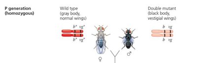
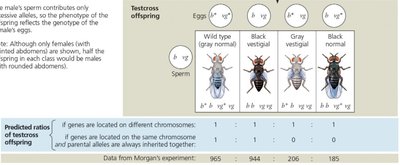
Thomas Hunt Morgan and Drosophila Genetics
Morgan's Experiments
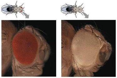
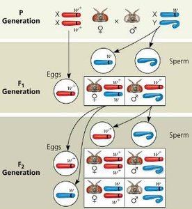
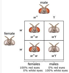
Thomas Hunt Morgan provided the first experimental evidence linking genes to chromosomes using the fruit fly Drosophila melanogaster. He studied mutations such as white eye color (w) versus wild-type red eyes (w+).
Sex-Linked Inheritance
Morgan discovered that the white-eye mutation was inherited in a pattern consistent with the X chromosome, providing evidence for sex-linked inheritance.
Autosomal vs. Sex-Linked Inheritance
Autosomal: Both sexes inherit traits equally.
Sex-linked (X-linked): Males (XY) express X-linked recessive traits more frequently because they have only one X chromosome.
Sex Chromosomes and Sex-Linked Genes
Structure and Function
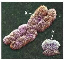
X chromosome: Large, contains many genes (about 1,100), not all related to sex determination.
Y chromosome: Small, few genes, mainly involved in male development.
Inheritance Patterns of Sex-Linked Genes
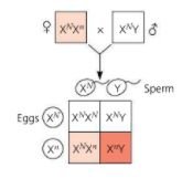
X-linked genes: Most sex-linked traits are X-linked. Males are hemizygous (only one X), so recessive alleles are always expressed if present.
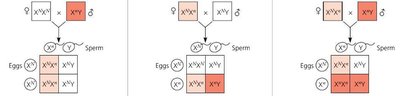
Carrier females (XDXd): Do not show the trait but can pass the allele to offspring.
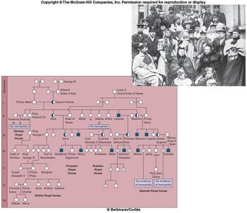
Examples: Color blindness, hemophilia.
Punnett Square Example: Hemophilia
Mother | Father | Daughters | Sons |
|---|---|---|---|
Carrier (XHXh) | Normal (XHY) | 50% carrier, 50% normal | 50% normal, 50% hemophiliac |
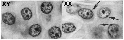
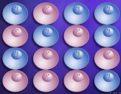
X Inactivation
Mechanism and Consequences
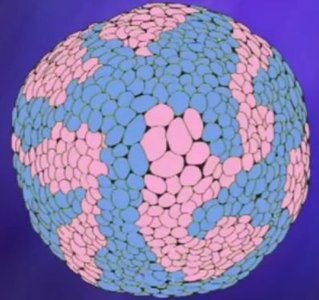
In female mammals, one X chromosome per cell is randomly inactivated during early embryogenesis, forming a Barr body. This ensures dosage compensation between males and females.
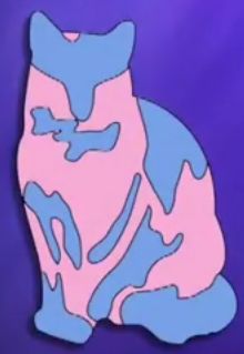
Example: Tortoiseshell Cats
Coat color genes are X-linked. Random X inactivation leads to patches of different colors in heterozygous females.
Linked Genes and Crossing Over
Definition and Behavior
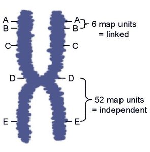
Linked genes: Genes located close together on the same chromosome tend to be inherited together.
Unlinked genes: Genes far apart on the same chromosome or on different chromosomes assort independently.
Crossing Over
During meiosis, homologous chromosomes exchange segments, producing recombinant chromosomes and increasing genetic diversity.
Linkage Maps
Linkage maps are constructed using recombination frequencies. The farther apart two genes are, the higher the probability that a crossover will occur between them.
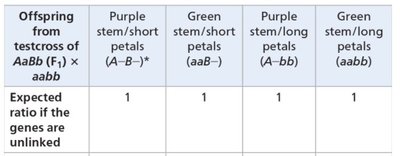
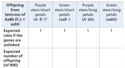
Testcross Example: Cosmos Plants
In a testcross between AaBb and aabb, if the genes are unlinked, the expected phenotypic ratio is 1:1:1:1. For 900 offspring, each phenotype is expected to appear 225 times.
Offspring phenotype | Expected number (of 900) |
|---|---|
Purple stem/short petals (A–B–) | 225 |
Green stem/short petals (aaB–) | 225 |
Purple stem/long petals (A–bb) | 225 |
Green stem/long petals (aabb) | 225 |
Chromosomal Alterations
Types of Structural Changes
Duplication: A segment of a chromosome is repeated. Can be beneficial or harmful (e.g., Huntington's disease).
Deletion: Loss of a chromosome segment. Can cause severe disorders (e.g., Cri du Chat syndrome, Williams syndrome).
Inversion: A chromosome segment is reversed. May cause infertility or disease if gene function is disrupted.
Translocation: A segment from one chromosome is transferred to another, often reciprocal. Can lead to cancer (e.g., Burkitt's lymphoma, chronic myelogenous leukemia).
Abnormal Chromosome Number
Nondisjunction and Aneuploidy
Nondisjunction: Failure of homologous chromosomes (anaphase I) or sister chromatids (anaphase II) to separate properly during meiosis, resulting in gametes with abnormal chromosome numbers.
Aneuploidy: Presence of an abnormal number of chromosomes (extra or missing).
Monosomy (2n-1): Missing a chromosome (e.g., Turner syndrome, XO).
Trisomy (2n+1): Extra chromosome (e.g., Down syndrome, trisomy 21).
Human Disorders Associated with Aneuploidy
Down Syndrome (Trisomy 21): Characterized by developmental delays, heart defects, and increased risk with maternal age.
Klinefelter Syndrome (XXY): Males with an extra X chromosome; sterile, some female characteristics.
XYY Syndrome: Males with an extra Y chromosome; usually taller, otherwise normal.
Turner Syndrome (XO): Females with only one X chromosome; short stature, sterile, underdeveloped secondary sex characteristics.
Triple X Syndrome (XXX): Females with an extra X chromosome; usually no significant symptoms due to X inactivation.
Summary Table: Types of Chromosomal Alterations
Alteration | Description | Example |
|---|---|---|
Duplication | Repeated segment | Huntington's disease (CAG repeats) |
Deletion | Missing segment | Cri du Chat (chromosome 5), Williams syndrome (chromosome 7) |
Inversion | Reversed segment | Acute myeloid leukemia (chromosome 16) |
Translocation | Segment moved to another chromosome | Burkitt's lymphoma (8 and 14), CML (9 and 22) |
Key Equations
Chi-square test for linkage:
Where is the observed number, is the expected number.
If , the difference is statistically significant (reject null hypothesis of independent assortment).
Conclusion
The chromosomal basis of inheritance provides a unifying explanation for Mendelian genetics, linking gene behavior to chromosome movement during meiosis. Understanding chromosomal alterations and their consequences is essential for interpreting patterns of inheritance and diagnosing genetic disorders.
