 Back
BackLEC 8:The Cytoskeleton: Structure and Function in Eukaryotic Cells
Study Guide - Smart Notes
 Tailored notes based on your materials, expanded with key definitions, examples, and context.
The Cytoskeleton in Eukaryotic Cells
Overview of the Cytoskeleton
The cytoskeleton is a dynamic network of protein filaments that provides structural support, organization, and motility to eukaryotic cells. It consists of three main types of protein filaments: microtubules, actin filaments (microfilaments), and intermediate filaments. Each type has distinct structures, functions, and associated proteins.
Microtubules: Thick, hollow tubes (25 nm diameter) made of tubulin proteins.
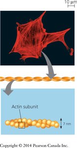
Actin filaments (microfilaments): Thin, flexible fibers (7 nm diameter) composed of actin subunits.
Intermediate filaments: Rope-like fibers (8–12 nm diameter) made of various proteins, providing mechanical strength.
Functions of the cytoskeleton:
Maintains cell shape and provides mechanical support.
Enables cell movement and changes in shape.
Acts as tracks for intracellular transport of organelles and vesicles.
Organizes the spatial arrangement of organelles within the cytoplasm.
Microtubules
Structure and Assembly
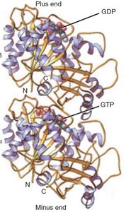
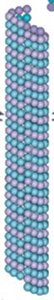
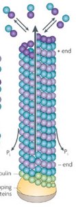
Microtubules are straight, hollow cylinders composed of repeating units of α-tubulin and β-tubulin proteins, which form heterodimers. These dimers assemble end-to-end to create protofilaments, and typically 13 protofilaments align side-by-side to form the microtubule wall.
Microtubules are polar structures with a plus end (fast-growing) and a minus end (slow-growing or anchored).
Assembly and disassembly occur primarily at the plus end.
Example: The dynamic instability of microtubules is essential for processes such as mitosis and intracellular transport.
Microtubule Dynamics
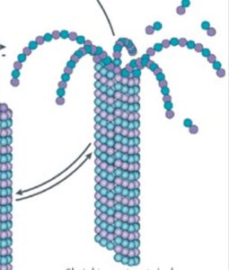
Microtubules exhibit dynamic instability, alternating between phases of growth and shrinkage. This behavior is regulated by the binding and hydrolysis of GTP on β-tubulin subunits.
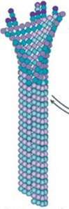
GTP-bound tubulin adds to the plus end, stabilizing the microtubule.
After incorporation, GTP is hydrolyzed to GDP, making the microtubule prone to depolymerization.
Catastrophe: Rapid shrinkage when the GTP cap is lost.
Rescue: Regrowth when new GTP-tubulin is added.

Microtubule Organizing Centers (MTOCs) and Centrosomes
In cells, the minus ends of microtubules are anchored in microtubule organizing centers (MTOCs). The primary MTOC in animal cells is the centrosome, which contains a pair of centrioles and a matrix of proteins, including γ-tubulin that nucleates microtubule growth.
γ-tubulin forms a ring complex that serves as a template for microtubule nucleation.
Centrioles are cylindrical structures composed of microtubule triplets, present in most animal cells but absent in plants and fungi.
Microtubule Motor Proteins
Microtubules serve as tracks for ATP-driven motor proteins, such as kinesin and dynein, which transport cargo (e.g., vesicles, organelles) along the microtubule network.
Kinesin generally moves toward the plus end (cell periphery).
Dynein moves toward the minus end (cell center).
Each step requires ATP hydrolysis:
Microtubules in Cell Division and Motility
During mitosis, microtubules reorganize to form the mitotic spindle, which separates chromosomes. Microtubules are also the main structural component of cilia and flagella, which are used for cell movement.
Cilia: Numerous, short, coordinated beating (e.g., in respiratory tract).
Flagella: Few, long, undulating motion (e.g., sperm tail).
Both structures have a "9+2" arrangement of microtubules and use dynein for movement.
Actin Filaments (Microfilaments)
Structure and Function
Actin filaments are thin, flexible fibers composed of actin protein subunits arranged in a helical chain. They are abundant in all eukaryotic cells and are especially important in muscle contraction, cell shape, and movement.
Actin filaments have polarity: a plus (barbed) end and a minus (pointed) end.
Growth occurs more rapidly at the plus end.
Actin interacts with myosin motor proteins to generate contractile forces.
Example: In muscle cells, actin and myosin filaments slide past each other to produce contraction.
Cellular Structures Formed by Actin
Microvilli: Increase surface area in epithelial cells (e.g., intestine).
Lamellipodia and filopodia: Protrusions for cell migration.
Contractile ring: Drives cytokinesis during cell division.
Contractile bundles: Provide mechanical support and contractility.
Intermediate Filaments
Structure and Function
Intermediate filaments are strong, rope-like fibers that provide mechanical strength to cells and tissues. Unlike microtubules and actin filaments, they are less dynamic and do not have polarity.
Composed of various proteins depending on cell type (e.g., keratins, vimentin, desmin, lamins).
Form stable, permanent structures in the cell.
Support the nuclear envelope (nuclear lamins) and maintain cell integrity under stress.
Example: Keratin filaments in epithelial cells provide resilience to mechanical stress.
Summary Table: Cytoskeletal Filaments
Filament Type | Diameter | Protein Subunits | Main Functions | Polarity | Dynamics |
|---|---|---|---|---|---|
Microtubules | 25 nm | α- and β-tubulin | Cell shape, transport, mitosis, cilia/flagella | Yes | Highly dynamic |
Actin Filaments | 7 nm | Actin | Cell shape, movement, muscle contraction | Yes | Dynamic |
Intermediate Filaments | 8–12 nm | Various (e.g., keratin, vimentin) | Mechanical strength, nuclear support | No | Stable |
