 Back
BackThe Cytoskeleton: Structure, Components, and Functions
Study Guide - Smart Notes
 Tailored notes based on your materials, expanded with key definitions, examples, and context.
Cytoskeleton
Overview and Functions
The cytoskeleton is a dynamic network of protein filaments and tubules that extends throughout the cytoplasm of eukaryotic cells. It provides structural support, enables cellular movement, facilitates intracellular transport, and regulates biochemical activities by transmitting mechanical forces.
Mechanical Support: Maintains cell shape and structural integrity.
Cell Motility: Enables movement of cells and cellular structures.
Intracellular Transport: Mediates movement of organelles and molecules within the cell.
Regulation: Transmits mechanical signals to regulate cellular activities.
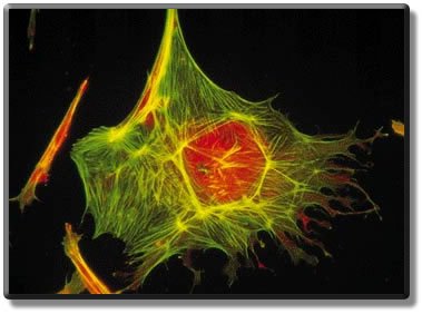
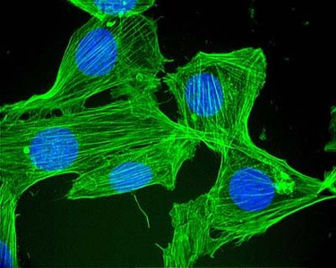
Cytoskeleton Components
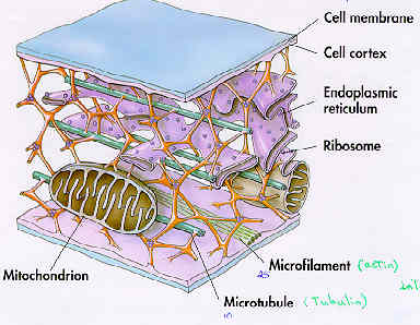
The cytoskeleton is composed of three main types of filaments:
Microtubules
Microfilaments (actin filaments)
Intermediate filaments
Microtubules
Structure and Assembly
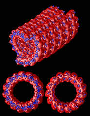
Microtubules are hollow tubes composed of tubulin protein dimers (α-tubulin and β-tubulin). These dimers polymerize to form protofilaments, which assemble into the cylindrical wall of the microtubule.
Outer diameter: ~25 nm
Lumen (inner diameter): ~15 nm
Length: 200 nm to 25,000 nm
Functions of Microtubules
Maintains cell shape: Resists compression forces.
Cell motility: Forms the structural basis of cilia and flagella.
Intracellular transport: Moves vesicles and organelles (e.g., from Golgi to plasma membrane).
Chromosome movement: Essential during cell division.
Microtubules grow and shrink by the addition or removal of tubulin dimers at their ends, a process known as dynamic instability.
Centrosomes and Centrioles
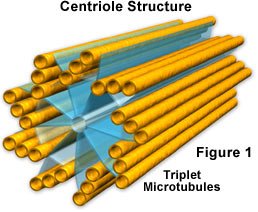
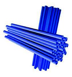
In animal cells, microtubules often originate from centrosomes, which contain two centrioles arranged at a 90° angle. Each centriole is composed of nine sets of triplet microtubules.
Centrioles: ~250 nm in diameter; divide during cell division.
Plant cells: Lack centrioles but still organize microtubules.
Cilia and Flagella
Structure and Function
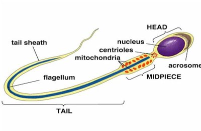
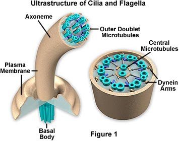
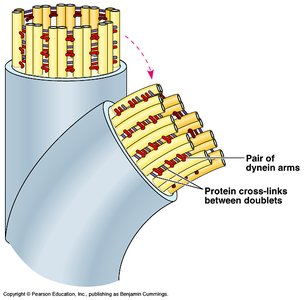
Cilia and flagella are cellular appendages that enable movement. Both are composed of microtubules arranged in a characteristic "9 + 2" pattern: nine doublets in a ring and two single microtubules in the center.
Cilia: Numerous, short, beat like oars (~0.25 µm diameter, 2–20 µm length, 40–60 strokes/sec).
Flagella: Usually single, longer, undulate (~0.25 µm diameter, 10–200 µm length).
Ultrastructure and Movement
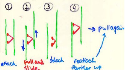
The movement of cilia and flagella is powered by dynein motor proteins, which use ATP to slide microtubule doublets against each other. This sliding causes bending and movement.
Dynein arms: Attach to adjacent doublets, pull, detach, and reattach further along, producing movement.
Radial spokes: Connect doublets to central microtubules.
Basal body: Anchors cilium/flagellum to the cell; structurally similar to a centriole.
Microfilaments (Actin Filaments)
Structure and Functions
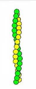
Microfilaments are thin, flexible filaments composed of actin protein. They are ~7 nm in diameter and consist of two intertwined strands of actin monomers.
Maintains cell shape: Resists tension (pulling forces).
Motility: Involved in muscle contraction, cell division, and cytoplasmic streaming.
Microfilaments in Muscle Contraction and Cell Movement
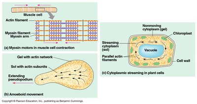
Microfilaments play a crucial role in muscle contraction, where actin filaments interact with myosin filaments. This sliding mechanism shortens the cell, resulting in contraction. Similar actin-myosin interactions drive amoeboid movement and cytoplasmic streaming in plant cells.
Gel state: High concentration of microfilaments makes cytoplasm rigid.
Sol state: Lower concentration allows cytoplasm to flow.
Cytoplasmic streaming: Circular motion of cytoplasm to distribute materials.
Microfilaments in Microvilli
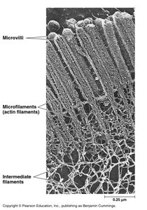
Microfilaments are present in the core of microvilli, which are cellular projections that increase surface area for absorption, especially in intestinal cells.
Summary Table: Cytoskeletal Components
Component | Structure | Diameter | Main Functions |
|---|---|---|---|
Microtubules | Hollow tubes of tubulin dimers | ~25 nm | Cell shape, motility, transport, chromosome movement |
Microfilaments | Two intertwined actin strands | ~7 nm | Cell shape, muscle contraction, cell division, streaming |
Intermediate filaments | Fibrous proteins coiled into cables | 8–12 nm | Cell shape, anchoring organelles |
Additional info: Intermediate filaments are less dynamic than microtubules and microfilaments and provide long-term structural stability.
