 Back
The Eukaryotic Cell Cycle, Mitosis, and Cancer
Study Guide - Smart Notes
 Tailored notes based on your materials, expanded with key definitions, examples, and context.
The Cell Cycle and Cellular Division
Introduction to Cellular Division
Cellular division is a fundamental process for organismal growth, development, and tissue repair. In eukaryotes, this process is tightly regulated to ensure the accurate transmission of genetic material. Abnormalities in cell division can lead to diseases such as cancer, characterized by uncontrolled cell proliferation.
Key Functions of Cell Division:
Growth of multicellular organisms
Repair and replacement of damaged cells
Asexual reproduction in some organisms
Types of Cell Division:
Mitosis: Division in eukaryotic cells producing genetically identical daughter cells
Binary Fission: Division in prokaryotic cells
The Eukaryotic Cell Cycle
Phases of the Cell Cycle
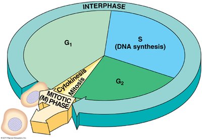
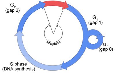
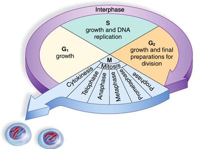
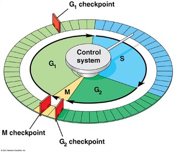
The eukaryotic cell cycle consists of a series of stages that prepare the cell for division and ensure the accurate duplication and segregation of genetic material. The main phases are interphase (G1, S, G2) and the mitotic (M) phase.
Interphase: Period of cell growth and DNA replication, subdivided into:
G1 phase (First Gap): Cell grows and carries out normal functions.
S phase (Synthesis): DNA is replicated.
G2 phase (Second Gap): Cell prepares for mitosis; additional growth and protein synthesis occur.
M phase (Mitotic phase): Includes mitosis (nuclear division) and cytokinesis (cytoplasmic division).
G0 Phase
Some cells exit the cell cycle and enter a quiescent state called G0, where they do not divide. Examples include most neurons and muscle cells in adults. Stem cells and some immune cells can re-enter the cycle from G0.
Overview of Mitosis and Cytokinesis
Mitosis is the process by which the nucleus divides, followed by cytokinesis, which divides the cytoplasm, resulting in two genetically identical daughter cells.
DNA Replication and Chromosome Structure
DNA Replication in S Phase
During the S phase, the cell duplicates its DNA, ensuring each daughter cell receives an identical set of chromosomes. DNA replication involves unwinding the double helix and synthesizing new complementary strands.
Major Steps in DNA Replication:
Unwinding of the DNA double helix
Synthesis of new DNA strands by DNA polymerase
Chromatin, Chromosomes, and Sister Chromatids
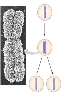
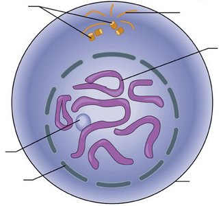
During interphase, DNA exists as chromatin, a less condensed form. Prior to mitosis, chromatin condenses into visible chromosomes, each consisting of two identical sister chromatids joined at a centromere.
Chromatin: DNA-protein complex in a relaxed state during interphase
Chromosome: Condensed DNA structure visible during mitosis
Sister Chromatids: Identical copies of a chromosome, joined at the centromere
Mitosis: Stages and Mechanisms
Stages of Mitosis
Mitosis is divided into several stages, each characterized by specific events involving chromosomes, the nuclear envelope, and the mitotic spindle.
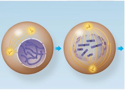
Prophase: Chromatin condenses into chromosomes; spindle apparatus begins to form; nuclear envelope starts to break down.
Prometaphase: Nuclear envelope fragments; spindle fibers attach to kinetochores on chromosomes.
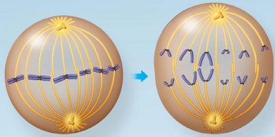
Metaphase: Chromosomes align at the metaphase plate (equatorial plane).
Anaphase: Sister chromatids are separated and pulled toward opposite poles.
Telophase: Nuclear envelopes reform around the separated chromosomes; chromosomes begin to decondense.
Mitotic Spindle and Chromosome Movement
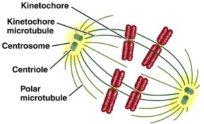
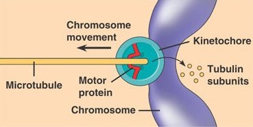
The mitotic spindle is composed of microtubules and associated proteins. It organizes and separates chromosomes during mitosis. Key components include centrosomes, centrioles, and kinetochores.
Centrosome: Microtubule-organizing center
Kinetochore: Protein structure on chromatids where spindle fibers attach
Microtubules: Dynamic filaments that pull chromatids apart
Cytokinesis
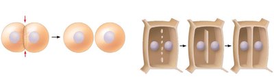
Cytokinesis is the division of the cytoplasm, resulting in two separate daughter cells. In animal cells, a cleavage furrow forms, while in plant cells, a cell plate develops to separate the cells.
Regulation of the Cell Cycle
Cell Cycle Checkpoints
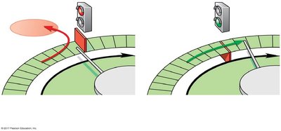
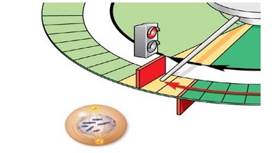
The cell cycle is regulated by checkpoints that ensure each phase is completed accurately before the next begins. Major checkpoints include G1, G2, and M checkpoints.
G1 Checkpoint: Determines if the cell will proceed with division or enter G0
G2 Checkpoint: Ensures DNA replication is complete and undamaged
M Checkpoint: Ensures all chromosomes are properly attached to the spindle before anaphase
Molecular Control: Cyclins and Cdks
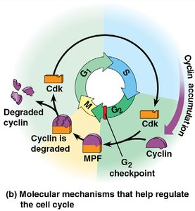
Protein kinases called cyclin-dependent kinases (Cdks) and their regulatory partners, cyclins, control the progression of the cell cycle. Cdk activity fluctuates with cyclin concentration, driving the cell through checkpoints.
MPF (Maturation Promoting Factor): A Cdk-cyclin complex that triggers the cell's passage from G2 to M phase
Mutations, Cancer, and Cell Cycle Dysregulation
Mutations and Their Consequences
Mutations are changes in the DNA sequence that can arise from errors during replication or from external mutagens (e.g., UV light, chemicals). While most mutations are harmless or repaired, some can disrupt cell cycle regulation and lead to cancer.
Types of Mutations:
Point mutations (substitutions, insertions, deletions)
Large-scale chromosomal rearrangements
Mutation Rates: DNA replication is highly accurate, with error rates as low as 1 in 109 to 1010 base pairs due to proofreading and repair mechanisms.
Mutagens and Types of Mutations
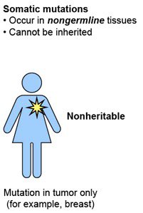
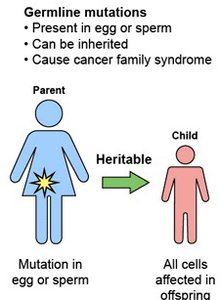
Mutagens are agents that cause mutations. Mutations can be classified as germline (inherited) or somatic (acquired in body cells).
Type | Description | Inheritance |
|---|---|---|
Germline Mutation | Occurs in egg or sperm; present in all cells of offspring | Heritable |
Somatic Mutation | Occurs in non-germline tissues; affects only some cells | Not heritable |
Cancer: Uncontrolled Cell Division
Cancer arises from the accumulation of mutations that disrupt normal cell cycle regulation, leading to uncontrolled cell growth and division. Malignant tumors can invade other tissues (metastasis) and evade immune detection.
Hallmarks of Cancer:
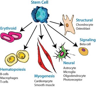
Dedifferentiation (loss of specialized function)
Immune system evasion
Metastasis (spread to other tissues)
Loss of cell cycle or contact inhibition control
Cancer and Aging
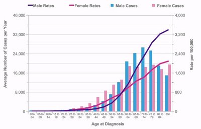
The incidence of cancer increases with age, reflecting the time required for multiple mutations to accumulate. Most adults acquire new mutations not inherited from their parents, contributing to cancer risk over time.
Key Vocabulary
Asexual reproduction: Offspring arise from a single parent without genetic recombination
Binary fission: Prokaryotic cell division
Centromere: Region joining sister chromatids
Centrosome: Organelle organizing microtubules
Chromatin: DNA-protein complex in non-dividing cells
Chromosome: Condensed DNA structure during division
Kinetochore: Protein complex on chromatids for spindle attachment
Mutation: Change in DNA sequence
Mutagen: Agent causing mutations
Differentiation: Process by which cells become specialized
