 Back
The Evolution of Multicellularity: Sponges and the Early Animal Radiation
Study Guide - Smart Notes
 Tailored notes based on your materials, expanded with key definitions, examples, and context.
Recap: The Origin of Life and Early Earth
Early Earth and the First Life Forms
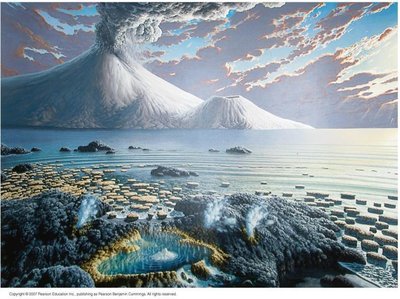
Understanding the conditions of early Earth is essential for studying the origin of life. The planet initially had extreme conditions, lacking oxygen, with a hot surface that eventually cooled to form a stable crust and pools of water. These pools enabled the spontaneous formation of organic compounds, setting the stage for the emergence of life.
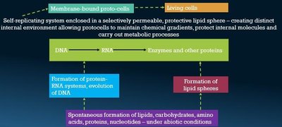
Abiotic Synthesis: Organic molecules formed spontaneously under physical conditions, leading to the creation of nucleic acids and protein-RNA systems.
Protocells: Lipid spheres surrounded nucleic acids, forming the first membrane-bound protocells capable of replication and basic metabolism.

Recap: The First Cells and the Evolution of Eukaryotes
Prokaryotes and the Rise of Eukaryotes
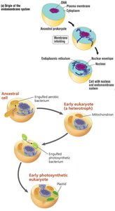
The earliest cells were prokaryotic, utilizing anaerobic pathways and existing as heterotrophs. The evolution of cyanobacteria introduced photosynthesis, increasing atmospheric oxygen and paving the way for aerobic eukaryotes. Eukaryotes arose through the development of internal membranes and endosymbiosis, leading to complex cellular structures.
Prokaryotes: Simple, anaerobic, and heterotrophic.
Cyanobacteria: Photosynthetic prokaryotes that produced oxygen.
Endosymbiosis: The origin of mitochondria and plastids in eukaryotes through symbiotic relationships with prokaryotes.
Kingdom Animalia and the Evolution of Multicellularity
Characteristics of Animals
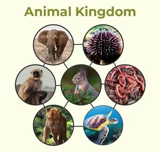
Animals are multicellular, diploid, heterotrophic eukaryotes that originated during the Precambrian era. They require oxygen, reproduce sexually or asexually, and exhibit motility at some life stage. Animal cells lack cell walls and are supported by an extracellular matrix composed of collagen and glycoproteins. Most animals develop from embryos with three primary germ layers: ectoderm, endoderm, and mesoderm (triploblastic), except for sponges and cnidarians.
Multicellularity: Many cells with coordinated metabolic activity and differentiation into tissues and organs.
Germ Layers: Ectoderm (outer), endoderm (inner), and mesoderm (middle; present in most animals).
Origin of Multicellularity
Hypotheses for the Evolution of Multicellularity
Multicellularity is defined by the presence of many cells with specialized functions. Two main hypotheses explain its origin:
Syncytial Theory: A multinucleated ciliate became compartmentalized into individual cells.
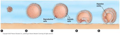
Colonial Theory (Dominant): Animals evolved from colonial flagellated protists, with cells becoming specialized and forming a division of labor.
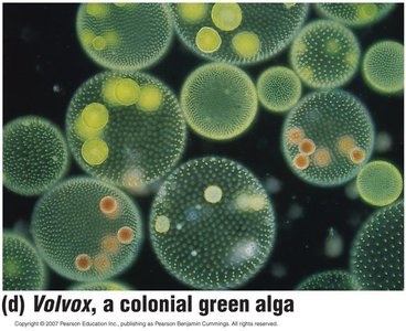
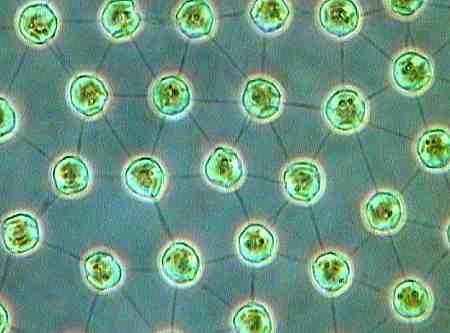
The colonial theory is supported by the observation of modern colonial protists, such as Volvox, which display cell specialization and division of labor.
Advantages of Multicellularity
Benefits of Being Multicellular
Multicellularity offers several evolutionary advantages:
Ability to grow larger in size
Improved predation and motility
Withstanding environmental forces
Differentiation of labor among cells
Specialization and interdependency
First Radiation: Sponges (Phylum Porifera)
Introduction to Sponges
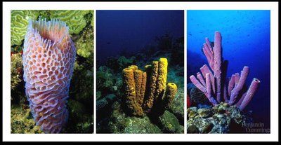
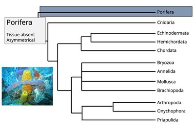
Sponges are the earliest diverging animal group, representing a living link between colonial protozoans and more complex animals. They exist at the cellular level of organization, lacking true tissues, organs, and symmetry. Sponges can reorganize themselves after being broken apart, demonstrating their primitive but adaptable body plan.
Habitat: Marine and freshwater environments
Body Plan: Asymmetrical, no true tissues or organs
Cell Types: Distinct cell types with division of labor
Phylogenetic Position and Evolutionary Significance
Sponges occupy the basal position in the animal tree of life, diverging before the evolution of true tissues and organs. Their ancestral cell type, the choanocyte, closely resembles choanoflagellates, linking them to unicellular ancestors. Fossil evidence dates sponges to 600–890 million years ago.
Feature | Sponges (Porifera) | Significance |
|---|---|---|
Cell structure | First multicellular animals | Oldest animal phylum – early radiation |
Ancestral cell type | Choanocytes | Link to unicellular ancestors |
Earliest fossil evidence | 600–890 mya | Dates the rise of animal multicellularity |
Body complexity | No true tissues/organs | Model for early animal organization |
Evolutionary status | Basal metazoan lineage | Insight into origin of complex animals |
Structure of Sponges
Basic Body Plan
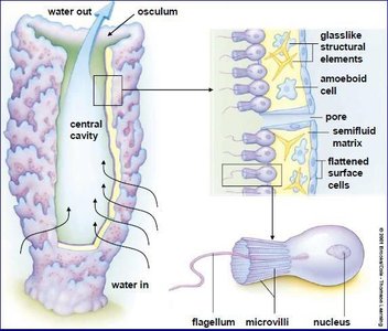
Sponges have a simple but functional structure optimized for water flow and filtration. Their bodies are typically organized into two or three layers:
Outer Layer: Pinacocytes (protective, mildly contractile cells), some modified into porocytes (ostia) for water regulation.
Mesohyl: Gelatinous matrix containing amoebocytes (secrete spicules, digest food, differentiate into other cells, and participate in asexual reproduction).
Inner Layer: Choanocytes (collar cells) generate water currents and capture food particles.
Canal System and Water Flow
Sponges feed by filtering water through a system of pores and canals:
Ostia: Inlet pores for water entry
Spongocoel: Central cavity where filtered water collects
Choanocytes: Line internal chambers, beat flagella to create water currents
Osculum: Outlet for water to exit the sponge
Defensive Systems in Sponges
Structural Defenses
Sponges possess a skeleton made of spicules (microscopic rods or needles of silica or calcium carbonate) embedded in the mesohyl. These spicules provide support and act as a physical barrier against predators, making the sponge tough and unpalatable.
Spicule Types: Megascleres (large, main framework), microscleres (small, scattered), monaxon (single axis), triaxon (three axes), tetraxon (four rays), polyaxon, and spheres.
Chemical Defenses
Sponges produce secondary metabolites (biotoxins, deterrents) that serve as antimicrobial agents and allelochemicals to prevent competition and predation.
Defence Mechanism | Description | Function |
|---|---|---|
Chemical defences | Secondary metabolites (biotoxins, deterrents) | Prevent microbial infection, deter predators, inhibit competitors |
Structural defences | Dense spicules | Mechanical barrier, reduce predation |
Life Cycle of Sponges
Sexual Reproduction
Sponges can reproduce sexually, typically as hermaphrodites (producing both eggs and sperm, but not simultaneously to avoid self-fertilization). Sperm are released into the water and carried to other sponges, where fertilization occurs. The resulting larva is free-swimming, allowing for dispersal before settling and metamorphosing into a sessile adult.
Asexual Reproduction
Asexual reproduction can occur via budding, fragmentation, or the formation of gemmules (resistant internal buds that survive harsh conditions). Offspring produced asexually are genetically identical to the parent.
Example: Sponges in coral reefs demonstrate both sexual and asexual reproduction, contributing to their resilience and ecological success.
