 Back
The Human Reproductive System: Structure, Function, and Regulation
Study Guide - Smart Notes
 Tailored notes based on your materials, expanded with key definitions, examples, and context.
The Human Reproductive System
Overview of Human Reproduction
The human reproductive system is responsible for producing, maintaining, and transporting gametes (sperm and eggs), as well as supporting fertilization, pregnancy, and the development of offspring. Both male and female systems have specialized structures and functions that ensure the continuation of the species.
Male Reproductive System
Major Functions
Produce and maintain sperm
Transport sperm and semen
Discharge sperm into the female reproductive tract
Produce testosterone
Anatomy of the Male Reproductive System
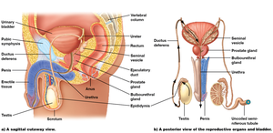
Testes: Produce sperm and testosterone. Located in the scrotum, which maintains a temperature slightly lower than body temperature for optimal sperm production.
Seminiferous tubules: Site of sperm production within the testes.
Epididymis: Sperm mature and are stored here.
Ductus (vas) deferens: Transports sperm from the epididymis to the ejaculatory duct.
Urethra: Conducts semen (and urine) to the outside of the body.
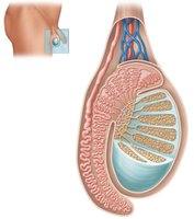
Route of Sperm Through Male Reproductive Structures
Seminiferous tubules → Epididymis → Ductus deferens → Ejaculatory duct → Urethra
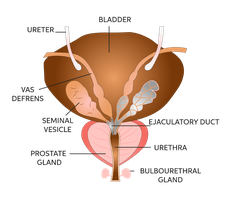
Accessory Glands
Accessory glands produce secretions that support sperm and form semen:
Seminal vesicles: Secrete fructose and most of the seminal fluid.
Prostate gland: Secretes a watery, alkaline fluid to raise vaginal pH.
Bulbourethral glands: Secrete lubricating mucus and cleanse the urethra.
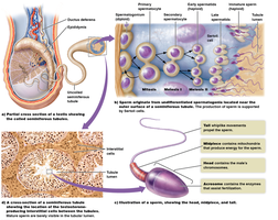
Sperm Production: Mitosis and Meiosis
Sperm are produced in the seminiferous tubules through a series of cell divisions:
Mitosis: Produces more spermatogonia (sperm precursor cells).
Meiosis: Reduces chromosome number by half, producing haploid sperm (n = 23).
Hormonal Regulation of Male Reproduction
Testosterone production and sperm formation are regulated by hormones:
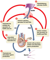
Gonadotropin-releasing hormone (GnRH): From the hypothalamus, stimulates the release of LH and FSH from the anterior pituitary.
Luteinizing hormone (LH): Stimulates Leydig cells to produce testosterone.
Follicle-stimulating hormone (FSH): Stimulates Sertoli cells, enhancing sperm formation.
Female Reproductive System
Major Functions
Produce and maintain ova (eggs)
Transport ova
Protect and nourish offspring until birth
Produce estrogen and progesterone
Anatomy of the Female Reproductive System
Ovaries: Release oocytes and secrete estrogen and progesterone.
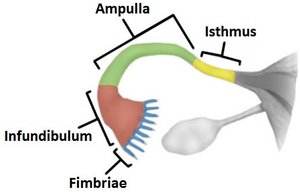
Oviducts (Fallopian tubes): Site of fertilization; transport oocyte to uterus.
Uterus: Site of embryo implantation and development.
Cervix: Lower opening of the uterus; allows passage of sperm and fetus.
Vagina: Canal for sexual intercourse and birth.
External genitalia (vulva): Includes labia majora, labia minora, and clitoris.
Ovarian Cycle
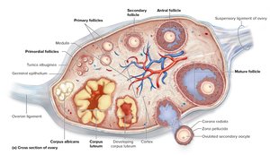
The ovarian cycle is a regular pattern of oocyte growth, maturation, and release:
At birth: ~1 million primary oocytes per ovary, arrested in meiosis I.
At puberty: About a dozen oocytes resume development each month; typically, one completes maturation and is ovulated every 28 days.
Ovulation: Release of a mature oocyte from the ovary (usually day 14 of the cycle).
Corpus luteum: Remnant of the follicle after ovulation; secretes hormones to support early pregnancy.
Uterine Cycle
The uterine (menstrual) cycle prepares the uterus for possible pregnancy:
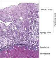
Menstrual phase (days 1–5): Estrogen and progesterone decrease; endometrial lining degenerates and is shed (menstruation).
Proliferative phase (days 6–14): Estrogen and progesterone increase; endometrial lining grows.
Ovulation (day 14): Oocyte is released; endometrium prepares for implantation.
Secretory phase (days 15–28): Corpus luteum produces hormones; endometrium is ready to accept a fertilized egg.
Hormonal Regulation of the Female Cycle
FSH and LH: Control the ovarian cycle (oocyte maturation and ovulation).
Estrogen and progesterone: Control the uterine cycle (endometrial changes).
Fertilization and Early Development
Fertilization occurs when a sperm unites with an egg, usually in the upper third of the oviduct, within 6–24 hours after intercourse. Only one sperm fertilizes the egg, forming a diploid zygote.
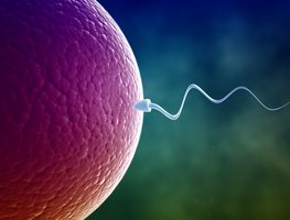
Barriers to sperm: Distance, acidic vaginal pH, cervical mucus, and white blood cells.
Twins: Identical (monozygotic) twins result from one fertilized oocyte splitting; fraternal (dizygotic) twins result from two oocytes fertilized by different sperm.
Pregnancy and Hormonal Changes
If fertilization occurs, embryonic tissue secretes human chorionic gonadotropin (hCG), which maintains the corpus luteum and its hormone production for 9–10 weeks, after which the placenta takes over.
High estrogen and progesterone levels prevent ovulation during pregnancy.
Pregnancy tests detect hCG in urine or blood.
If no fertilization occurs, the corpus luteum degenerates, hormone levels fall, and the cycle restarts.
Mammary Glands and Lactation
Mammary glands, located in the breasts, are modified sweat glands that produce milk to nourish the infant. They develop at puberty under the influence of estrogen and are part of the integumentary system.
Summary Table: Key Differences Between Male and Female Reproductive Systems
Feature | Male | Female |
|---|---|---|
Gamete Produced | Sperm (continuously after puberty) | Ova (one per cycle, from puberty to menopause) |
Primary Hormones | Testosterone | Estrogen, Progesterone |
Site of Gamete Production | Testes (seminiferous tubules) | Ovaries (follicles) |
Fertilization Site | Delivers sperm to female tract | Oviduct (Fallopian tube) |
Support for Offspring | None (beyond sperm delivery) | Uterus (pregnancy), Mammary glands (lactation) |
